 Movie
Movie Controller

[English] 日本語
 Yorodumi
- PDB-1hv9: STRUCTURE OF E. COLI GLMU: ANALYSIS OF PYROPHOSPHORYLASE AND ACET... -
+ Open data

- Basic information
| Entry | Database: PDB / ID: 1hv9 | ||||||
|---|---|---|---|---|---|---|---|
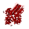
| Title | STRUCTURE OF E. COLI GLMU: ANALYSIS OF PYROPHOSPHORYLASE AND ACETYLTRANSFERASE ACTIVE SITES | ||||||
 Components Components | UDP-N-ACETYLGLUCOSAMINE PYROPHOSPHORYLASE | ||||||
 Keywords Keywords | TRANSFERASE / left-handed parallel beta-helix | ||||||
| Function / homology |  Function and homology information Function and homology informationglucosamine-1-phosphate N-acetyltransferase / glucosamine-1-phosphate N-acetyltransferase activity / UDP-N-acetylglucosamine diphosphorylase / UDP-N-acetylglucosamine diphosphorylase activity / UDP-N-acetylglucosamine biosynthetic process / lipid A biosynthetic process / peptidoglycan biosynthetic process / cell wall organization / cell morphogenesis / regulation of cell shape ...glucosamine-1-phosphate N-acetyltransferase / glucosamine-1-phosphate N-acetyltransferase activity / UDP-N-acetylglucosamine diphosphorylase / UDP-N-acetylglucosamine diphosphorylase activity / UDP-N-acetylglucosamine biosynthetic process / lipid A biosynthetic process / peptidoglycan biosynthetic process / cell wall organization / cell morphogenesis / regulation of cell shape / magnesium ion binding / membrane / identical protein binding / cytosol Similarity search - Function | ||||||
| Biological species |  | ||||||
| Method |  X-RAY DIFFRACTION / MIR / Resolution: 2.1 Å X-RAY DIFFRACTION / MIR / Resolution: 2.1 Å | ||||||
 Authors Authors | Olsen, L.R. / Roderick, S.L. | ||||||
 Citation Citation | Journal: Biochemistry / Year: 2001 Title: Structure of the Escherichia coli GlmU pyrophosphorylase and acetyltransferase active sites. Authors: Olsen, L.R. / Roderick, S.L. #1: Journal: Acta Crystallogr.,Sect.D / Year: 2001Title: Purification, Crystallization and Preliminary X-ray Data for Escherichia coli GlmU: A Bifunctional Acetyltransferase/Uridyltransferase Authors: Olsen, L.R. / Tian, Y. / Roderick, S.L. | ||||||
| History |
|
- Structure visualization
| Structure viewer | Molecule: MolmilJmol/JSmol |
|---|
- Downloads & links
-Download
| PDBx/mmCIF format | 1hv9.cif.gz | 191.6 KB | Display | PDBx/mmCIF format |
|---|---|---|---|---|
| PDB format | pdb1hv9.ent.gz | 150.5 KB | Display | PDB format |
| PDBx/mmJSON format | 1hv9.json.gz | Tree view | PDBx/mmJSON format | |
| Others |  Other downloads Other downloads |
-Validation report
| Arichive directory | https://data.pdbj.org/pub/pdb/validation_reports/hv/1hv9ftp://data.pdbj.org/pub/pdb/validation_reports/hv/1hv9 | HTTPS FTP |
|---|
-Related structure data
| Similar structure data |
|---|


-Links
 PDBj
PDBj


- Assembly
| Deposited unit | 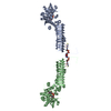
| |||||||||||||||
|---|---|---|---|---|---|---|---|---|---|---|---|---|---|---|---|---|
| 1 | 
| |||||||||||||||
| 2 | 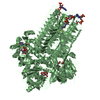
| |||||||||||||||
| 3 | 
| |||||||||||||||
| Unit cell |
| |||||||||||||||
| Components on special symmetry positions |
|
-Components
| #1: Protein | Mass: 49250.906 Da / Num. of mol.: 2 Source method: isolated from a genetically manipulated source Source: (gene. exp.) References: UniProt: P0ACC7, UDP-N-acetylglucosamine diphosphorylase #2: Chemical | ChemComp-CO /   Mass: 58.933 Da / Num. of mol.: 5 / Source method: obtained synthetically / Formula: Co Mass: 58.933 Da / Num. of mol.: 5 / Source method: obtained synthetically / Formula: Co#3: Chemical |   Mass: 767.534 Da / Num. of mol.: 2 / Source method: obtained synthetically / Formula: C21H36N7O16P3S Mass: 767.534 Da / Num. of mol.: 2 / Source method: obtained synthetically / Formula: C21H36N7O16P3S#4: Chemical |   Mass: 607.354 Da / Num. of mol.: 2 / Source method: obtained synthetically / Formula: C17H27N3O17P2 Mass: 607.354 Da / Num. of mol.: 2 / Source method: obtained synthetically / Formula: C17H27N3O17P2#5: Water | ChemComp-HOH / |  Mass: 18.015 Da / Num. of mol.: 257 / Source method: isolated from a natural source / Formula: H2O Mass: 18.015 Da / Num. of mol.: 257 / Source method: isolated from a natural source / Formula: H2O |
|---|
-Experimental details
-Experiment
| Experiment | Method: X-RAY DIFFRACTION / Number of used crystals: 2 |
|---|
- Sample preparation
| Crystal | Density Matthews: 3.46 Å3/Da / Density % sol: 64.41 % | ||||||||||||||||||||||||||||||||||||||||||||||||
|---|---|---|---|---|---|---|---|---|---|---|---|---|---|---|---|---|---|---|---|---|---|---|---|---|---|---|---|---|---|---|---|---|---|---|---|---|---|---|---|---|---|---|---|---|---|---|---|---|---|
| Crystal grow | Temperature: 293 K / Method: vapor diffusion, hanging drop / pH: 6.4 Details: MES, ammonium sulfate, magnesium chloride, cobalt chloride, pH 6.4, VAPOR DIFFUSION, HANGING DROP, temperature 293.0K | ||||||||||||||||||||||||||||||||||||||||||||||||
| Crystal grow | *PLUS PH range low: 6.4 / PH range high: 5.4 | ||||||||||||||||||||||||||||||||||||||||||||||||
| Components of the solutions | *PLUS
|
-Data collection
| Diffraction | Mean temperature: 293 K |
|---|---|
| Diffraction source | Source: ROTATING ANODE / Type: RIGAKU / Wavelength: 1.5418 Å |
| Detector | Type: RIGAKU RAXIS IV / Detector: IMAGE PLATE / Date: Apr 6, 1999 / Details: confocal mirrors |
| Radiation | Monochromator: confocal mirrors / Protocol: SINGLE WAVELENGTH / Monochromatic (M) / Laue (L): M / Scattering type: x-ray |
| Radiation wavelength | Wavelength: 1.5418 Å / Relative weight: 1 |
| Reflection | Resolution: 2.1→30.25 Å / Num. all: 391782 / Num. obs: 391782 / % possible obs: 96.4 % / Observed criterion σ(F): 0 / Observed criterion σ(I): -3 / Redundancy: 5 % / Biso Wilson estimate: 9.5 Å2 / Rmerge(I) obs: 0.107 / Net I/σ(I): 9.7 |
| Reflection shell | Resolution: 2.1→2.18 Å / Redundancy: 2.1 % / Rmerge(I) obs: 0.233 / Mean I/σ(I) obs: 1.62 / % possible all: 82.2 |
| Reflection | *PLUS Num. obs: 91622 / % possible obs: 98 % / Num. measured all: 495967 / Rmerge(I) obs: 0.136 |
- Processing
| Software |
| ||||||||||||||||||||||||||||||||||||||||
|---|---|---|---|---|---|---|---|---|---|---|---|---|---|---|---|---|---|---|---|---|---|---|---|---|---|---|---|---|---|---|---|---|---|---|---|---|---|---|---|---|---|
| Refinement | Method to determine structure: MIR / Resolution: 2.1→30.25 Å / Rfactor Rfree error: 0.004 / Data cutoff high absF: 10000000 / Data cutoff low absF: 0 / Isotropic thermal model: RESTRAINED / Cross valid method: THROUGHOUT / σ(F): 0 / σ(I): 0 / Stereochemistry target values: Engh & Huber
| ||||||||||||||||||||||||||||||||||||||||
| Displacement parameters | Biso mean: 21.6 Å2
| ||||||||||||||||||||||||||||||||||||||||
| Refine analyze |
| ||||||||||||||||||||||||||||||||||||||||
| Refinement step | Cycle: LAST / Resolution: 2.1→30.25 Å
| ||||||||||||||||||||||||||||||||||||||||
| Refine LS restraints |
| ||||||||||||||||||||||||||||||||||||||||
| LS refinement shell | Resolution: 2.1→2.18 Å / Rfactor Rfree error: 0.019 / Total num. of bins used: 10
| ||||||||||||||||||||||||||||||||||||||||
| Xplor file |
| ||||||||||||||||||||||||||||||||||||||||
| Software | *PLUS Name: X-PLOR(ONLINE) / Version: 3.851 / Classification: refinement | ||||||||||||||||||||||||||||||||||||||||
| Refinement | *PLUS σ(F): 0 / % reflection Rfree: 5 % | ||||||||||||||||||||||||||||||||||||||||
| Solvent computation | *PLUS | ||||||||||||||||||||||||||||||||||||||||
| Displacement parameters | *PLUS Biso mean: 21.6 Å2 | ||||||||||||||||||||||||||||||||||||||||
| Refine LS restraints | *PLUS
| ||||||||||||||||||||||||||||||||||||||||
| LS refinement shell | *PLUS Rfactor Rfree: 0.329 / % reflection Rfree: 4.9 % / Rfactor Rwork: 0.28 / Rfactor obs: 0.277 |
