 Movie
Movie Controller

[English] 日本語
 Yorodumi
- PDB-1v6t: Crystal Structure of Lactam Utilization Protein from Pyrococcus h... -
+ Open data

- Basic information
| Entry | Database: PDB / ID: 1v6t | ||||||
|---|---|---|---|---|---|---|---|
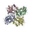
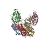
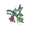
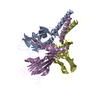
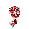
| Title | Crystal Structure of Lactam Utilization Protein from Pyrococcus horikoshii Ot3 | ||||||
 Components Components | Hypothetical UPF0271 protein PH0986 | ||||||
 Keywords Keywords | STRUCTURAL GENOMICS / UNKNOWN FUNCTION / TIM-BARREL / Lactam Utilization Protein / RIKEN Structural Genomics/Proteomics Initiative / RSGI | ||||||
| Function / homology |  Function and homology information Function and homology information5-oxoprolinase (ATP-hydrolysing) / 5-oxoprolinase (ATP-hydrolyzing) activity / carbohydrate metabolic process / ATP binding Similarity search - Function | ||||||
| Biological species |   Pyrococcus horikoshii (archaea) Pyrococcus horikoshii (archaea) | ||||||
| Method |  X-RAY DIFFRACTION / SYNCHROTRON / MAD / Resolution: 1.7 Å X-RAY DIFFRACTION / SYNCHROTRON / MAD / Resolution: 1.7 Å | ||||||
 Authors Authors | Mizutani, H. / Kunishima, N. / RIKEN Structural Genomics/Proteomics Initiative (RSGI) | ||||||
 Citation Citation | Journal: To be Published Title: Crystal Structure of Lactam Utilization Protein from Pyrococcus horikoshii Ot3 Authors: Mizutani, H. / Kunishima, N. | ||||||
| History |
|
- Structure visualization
| Structure viewer | Molecule: MolmilJmol/JSmol |
|---|
- Downloads & links
-Download
| PDBx/mmCIF format | 1v6t.cif.gz | 63.5 KB | Display | PDBx/mmCIF format |
|---|---|---|---|---|
| PDB format | pdb1v6t.ent.gz | 46.4 KB | Display | PDB format |
| PDBx/mmJSON format | 1v6t.json.gz | Tree view | PDBx/mmJSON format | |
| Others |  Other downloads Other downloads |
-Validation report
| Arichive directory | https://data.pdbj.org/pub/pdb/validation_reports/v6/1v6tftp://data.pdbj.org/pub/pdb/validation_reports/v6/1v6t | HTTPS FTP |
|---|
-Related structure data
| Similar structure data | |
|---|---|
| Other databases |

-Links
 PDBj
PDBj
- Assembly
| Deposited unit | 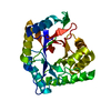
| ||||||||
|---|---|---|---|---|---|---|---|---|---|
| 1 | 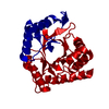
| ||||||||
| Unit cell |
| ||||||||
| Details | The biological assembly is a tetramer generated by the operations: 1-x,1-y,z, x,1-y,-z and 1-x,y,-z. |
-Components
| #1: Protein | Mass: 28469.713 Da / Num. of mol.: 1 Source method: isolated from a genetically manipulated source Source: (gene. exp.) Pyrococcus horikoshii (archaea) / Strain: OT3 / Plasmid: pET11a / Production host:  |
|---|---|
| #2: Water | ChemComp-HOH /  Mass: 18.015 Da / Num. of mol.: 177 / Source method: isolated from a natural source / Formula: H2O Mass: 18.015 Da / Num. of mol.: 177 / Source method: isolated from a natural source / Formula: H2O |
-Experimental details
-Experiment
| Experiment | Method: X-RAY DIFFRACTION / Number of used crystals: 1 |
|---|
- Sample preparation
| Crystal | Density Matthews: 2.35 Å3/Da / Density % sol: 47.24 % |
|---|---|
| Crystal grow | Temperature: 295 K / Method: microbatch / pH: 4.2 Details: sodium chloride, acetate, pH 4.2, microbatch, temperature 295K |
-Data collection
| Diffraction | Mean temperature: 100 K | ||||||||||||
|---|---|---|---|---|---|---|---|---|---|---|---|---|---|
| Diffraction source | Source: SYNCHROTRON / Site: SPring-8  / Beamline: BL26B1 / Wavelength: 1.00, 0.97914, 0.97964 / Beamline: BL26B1 / Wavelength: 1.00, 0.97914, 0.97964 | ||||||||||||
| Detector | Type: RIGAKU RAXIS V / Detector: IMAGE PLATE / Date: Oct 18, 2003 / Details: mirrors | ||||||||||||
| Radiation | Monochromator: MIRRORS / Protocol: MAD / Monochromatic (M) / Laue (L): M / Scattering type: x-ray | ||||||||||||
| Radiation wavelength |
| ||||||||||||
| Reflection | Resolution: 1.7→30 Å / Num. all: 28634 / Num. obs: 28634 / % possible obs: 100 % / Observed criterion σ(F): 0 / Observed criterion σ(I): 0 / Redundancy: 5.9 % / Biso Wilson estimate: 17.9 Å2 / Rmerge(I) obs: 0.043 / Rsym value: 0.04 / Net I/σ(I): 20.1 | ||||||||||||
| Reflection shell | Resolution: 1.7→1.76 Å / Redundancy: 5.14 % / Rmerge(I) obs: 0.259 / Mean I/σ(I) obs: 6.35 / Num. unique all: 2822 / Rsym value: 0.232 / % possible all: 100 |
- Processing
| Software |
| ||||||||||||||||||||||||||||||||||||||||||||||||||||||||||||
|---|---|---|---|---|---|---|---|---|---|---|---|---|---|---|---|---|---|---|---|---|---|---|---|---|---|---|---|---|---|---|---|---|---|---|---|---|---|---|---|---|---|---|---|---|---|---|---|---|---|---|---|---|---|---|---|---|---|---|---|---|---|
| Refinement | Method to determine structure: MAD / Resolution: 1.7→28.78 Å / Rfactor Rfree error: 0.006 / Data cutoff high absF: 1854240.53 / Data cutoff low absF: 0 / Isotropic thermal model: RESTRAINED / Cross valid method: THROUGHOUT / σ(F): 0 / Stereochemistry target values: Engh & Huber
| ||||||||||||||||||||||||||||||||||||||||||||||||||||||||||||
| Solvent computation | Solvent model: FLAT MODEL / Bsol: 46.696 Å2 / ksol: 0.364966 e/Å3 | ||||||||||||||||||||||||||||||||||||||||||||||||||||||||||||
| Displacement parameters | Biso mean: 22.1 Å2
| ||||||||||||||||||||||||||||||||||||||||||||||||||||||||||||
| Refine analyze |
| ||||||||||||||||||||||||||||||||||||||||||||||||||||||||||||
| Refinement step | Cycle: LAST / Resolution: 1.7→28.78 Å
| ||||||||||||||||||||||||||||||||||||||||||||||||||||||||||||
| Refine LS restraints |
| ||||||||||||||||||||||||||||||||||||||||||||||||||||||||||||
| LS refinement shell | Resolution: 1.7→1.76 Å / Rfactor Rfree error: 0.024 / Total num. of bins used: 10
| ||||||||||||||||||||||||||||||||||||||||||||||||||||||||||||
| Xplor file |
|
