 Movie
Movie Controller

[English] 日本語
 Yorodumi
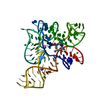
- PDB-3gx3: Crystal structure of the T. tengcongensis SAM-I riboswitch varian... -
+ Open data

- Basic information
| Entry | Database: PDB / ID: 3gx3 | ||||||
|---|---|---|---|---|---|---|---|
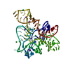
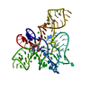
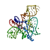
| Title | Crystal structure of the T. tengcongensis SAM-I riboswitch variant U34C/A94G bound with SAH | ||||||
 Components Components | RNA (94-MER) | ||||||
 Keywords Keywords | RNA / kink-turn / four-way junction / pseudoknot / riboswitch | ||||||
| Function / homology | : / S-ADENOSYL-L-HOMOCYSTEINE / RNA / RNA (> 10) Function and homology information Function and homology information | ||||||
| Method |  X-RAY DIFFRACTION / MOLECULAR REPLACEMENT / Resolution: 2.7 Å X-RAY DIFFRACTION / MOLECULAR REPLACEMENT / Resolution: 2.7 Å | ||||||
 Authors Authors | Montange, R.K. / Batey, R.T. | ||||||
 Citation Citation | Journal: J.Mol.Biol. / Year: 2010 Title: Discrimination between Closely Related Cellular Metabolites by the SAM-I Riboswitch. Authors: Montange, R.K. / Mondragon, E. / van Tyne, D. / Garst, A.D. / Ceres, P. / Batey, R.T. | ||||||
| History |
|
- Structure visualization
| Structure viewer | Molecule: MolmilJmol/JSmol |
|---|
- Downloads & links
-Download
| PDBx/mmCIF format | 3gx3.cif.gz | 63.7 KB | Display | PDBx/mmCIF format |
|---|---|---|---|---|
| PDB format | pdb3gx3.ent.gz | 46.5 KB | Display | PDB format |
| PDBx/mmJSON format | 3gx3.json.gz | Tree view | PDBx/mmJSON format | |
| Others |  Other downloads Other downloads |
-Validation report
| Arichive directory | https://data.pdbj.org/pub/pdb/validation_reports/gx/3gx3ftp://data.pdbj.org/pub/pdb/validation_reports/gx/3gx3 | HTTPS FTP |
|---|
-Related structure data
| Related structure data | 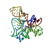 3gx2C 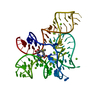 3gx5C 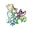 3gx6C 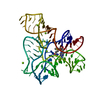 3gx7C 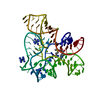 2gisS C: citing same article ( S: Starting model for refinement |
|---|---|
| Similar structure data |


-Links
 PDBj
PDBj


- Assembly
| Deposited unit | 
| ||||||||
|---|---|---|---|---|---|---|---|---|---|
| 1 |
| ||||||||
| Unit cell |
|
-Components
| #1: RNA chain | Mass: 30568.338 Da / Num. of mol.: 1 / Mutation: U34C, A94G / Source method: obtained synthetically Details: the sequence is based on a naturally occurring sequence found in T. tengcongensis | ||||
|---|---|---|---|---|---|
| #2: Chemical | ChemComp-SAH /   Type: L-peptide linking / Mass: 384.411 Da / Num. of mol.: 1 / Source method: obtained synthetically / Formula: C14H20N6O5S Type: L-peptide linking / Mass: 384.411 Da / Num. of mol.: 1 / Source method: obtained synthetically / Formula: C14H20N6O5S | ||||
| #3: Chemical | ChemComp-MG /   Mass: 24.305 Da / Num. of mol.: 9 / Source method: obtained synthetically / Formula: Mg Mass: 24.305 Da / Num. of mol.: 9 / Source method: obtained synthetically / Formula: Mg#4: Chemical | ChemComp-K / |   Mass: 39.098 Da / Num. of mol.: 1 / Source method: obtained synthetically / Formula: K Mass: 39.098 Da / Num. of mol.: 1 / Source method: obtained synthetically / Formula: K#5: Water | ChemComp-HOH / |  Mass: 18.015 Da / Num. of mol.: 12 / Source method: isolated from a natural source / Formula: H2O Mass: 18.015 Da / Num. of mol.: 12 / Source method: isolated from a natural source / Formula: H2O |
-Experimental details
-Experiment
| Experiment | Method: X-RAY DIFFRACTION / Number of used crystals: 1 |
|---|
- Sample preparation
| Crystal | Density Matthews: 2.52 Å3/Da / Density % sol: 51.22 % | ||||||||||||||||||||||||||||||||||||||||||||
|---|---|---|---|---|---|---|---|---|---|---|---|---|---|---|---|---|---|---|---|---|---|---|---|---|---|---|---|---|---|---|---|---|---|---|---|---|---|---|---|---|---|---|---|---|---|
| Crystal grow | Method: vapor diffusion, hanging drop / pH: 7 Details: 20 mM magnesium chloride, 80 mM potassium chloride, 12 mM spermine, 10% MPD, 40 mM cacodylate, pH 7.0, VAPOR DIFFUSION, HANGING DROP | ||||||||||||||||||||||||||||||||||||||||||||
| Components of the solutions |
|
-Data collection
| Diffraction | Mean temperature: 100 K |
|---|---|
| Diffraction source | Source: ROTATING ANODE / Type: RIGAKU RUH2R / Wavelength: 1.54 Å |
| Detector | Type: RIGAKU RAXIS IV++ / Detector: IMAGE PLATE / Date: Jan 19, 2009 / Details: osmic mirror |
| Radiation | Monochromator: Ni filter / Protocol: SINGLE WAVELENGTH / Monochromatic (M) / Laue (L): M / Scattering type: x-ray |
| Radiation wavelength | Wavelength: 1.54 Å / Relative weight: 1 |
| Reflection | Resolution: 2.7→20 Å / Num. obs: 8993 / % possible obs: 98.3 % / Observed criterion σ(I): 2 / Redundancy: 6.74 % / Rmerge(I) obs: 0.075 / Net I/σ(I): 15.7 |
| Reflection shell | Resolution: 2.7→2.8 Å / Redundancy: 7.06 % / Rmerge(I) obs: 0.369 / Mean I/σ(I) obs: 4.6 / % possible all: 97.6 |
- Processing
| Software |
| |||||||||||||||||||||||||||||||||||||||||||||||||
|---|---|---|---|---|---|---|---|---|---|---|---|---|---|---|---|---|---|---|---|---|---|---|---|---|---|---|---|---|---|---|---|---|---|---|---|---|---|---|---|---|---|---|---|---|---|---|---|---|---|---|
| Refinement | Method to determine structure: MOLECULAR REPLACEMENT Starting model: pdb entry 2GIS Resolution: 2.7→19.741 Å / SU ML: 0.54 / σ(F): 2.2 / Phase error: 33.49 / Stereochemistry target values: ML
| |||||||||||||||||||||||||||||||||||||||||||||||||
| Solvent computation | Shrinkage radii: 0.9 Å / VDW probe radii: 1.11 Å / Solvent model: FLAT BULK SOLVENT MODEL / Bsol: 23.38 Å2 / ksol: 0.245 e/Å3 | |||||||||||||||||||||||||||||||||||||||||||||||||
| Displacement parameters | Biso mean: 54.6 Å2 | |||||||||||||||||||||||||||||||||||||||||||||||||
| Refinement step | Cycle: LAST / Resolution: 2.7→19.741 Å
| |||||||||||||||||||||||||||||||||||||||||||||||||
| Refine LS restraints |
| |||||||||||||||||||||||||||||||||||||||||||||||||
| LS refinement shell |
|
