[English] 日本語
Yorodumi
- PDB-2ydh: Crystal structure of the SAM-I riboswitch A94G U34 G18U G19U vari... -

+
Open data


ID or keywords:

Loading...

-
Basic information

Entry
Database: PDB / ID: 2ydh
TitleCrystal structure of the SAM-I riboswitch A94G U34 G18U G19U variant in complex with SAM
ComponentsSAM-I RIBOSWITCH
KeywordsRNA / K-TURN
Function / homology: / S-ADENOSYLMETHIONINE / RNA / RNA (> 10)
Function and homology information
Biological speciesTHERMOANAEROBACTER TENGCONGENSIS (bacteria)
MethodX-RAY DIFFRACTION / SYNCHROTRON / MOLECULAR REPLACEMENT / Resolution: 2.9 Å
AuthorsSchroeder, K.T. / Daldrop, P. / Lilley, D.M.J.
CitationJournal: Structure / Year: 2011
Title: RNA Tertiary Interactions in a Riboswitch Stabilize the Structure of a Kink Turn.
Authors: Schroeder, K.T. / Daldrop, P. / Lilley, D.M.J.
History
DepositionMar 21, 2011Deposition site: PDBE / Processing site: PDBE
Revision 1.0Sep 7, 2011Provider: repository / Type: Initial release
Revision 1.1Sep 21, 2011Group: Database references
Revision 1.2Sep 28, 2016Group: Structure summary
Revision 1.3Dec 20, 2023Group: Data collection / Database references ...Data collection / Database references / Derived calculations / Other / Refinement description
Category: chem_comp_atom / chem_comp_bond ...chem_comp_atom / chem_comp_bond / database_2 / pdbx_database_status / pdbx_initial_refinement_model / pdbx_struct_conn_angle / struct_conn / struct_site
Item: _database_2.pdbx_DOI / _database_2.pdbx_database_accession ..._database_2.pdbx_DOI / _database_2.pdbx_database_accession / _pdbx_database_status.status_code_sf / _pdbx_struct_conn_angle.ptnr1_auth_comp_id / _pdbx_struct_conn_angle.ptnr1_auth_seq_id / _pdbx_struct_conn_angle.ptnr1_label_atom_id / _pdbx_struct_conn_angle.ptnr1_label_comp_id / _pdbx_struct_conn_angle.ptnr1_label_seq_id / _pdbx_struct_conn_angle.ptnr2_auth_seq_id / _pdbx_struct_conn_angle.ptnr2_label_asym_id / _pdbx_struct_conn_angle.ptnr3_auth_comp_id / _pdbx_struct_conn_angle.ptnr3_auth_seq_id / _pdbx_struct_conn_angle.ptnr3_label_atom_id / _pdbx_struct_conn_angle.ptnr3_label_comp_id / _pdbx_struct_conn_angle.ptnr3_label_seq_id / _pdbx_struct_conn_angle.value / _struct_conn.pdbx_dist_value / _struct_conn.ptnr1_auth_comp_id / _struct_conn.ptnr1_auth_seq_id / _struct_conn.ptnr1_label_asym_id / _struct_conn.ptnr1_label_atom_id / _struct_conn.ptnr1_label_comp_id / _struct_conn.ptnr1_label_seq_id / _struct_conn.ptnr2_auth_comp_id / _struct_conn.ptnr2_auth_seq_id / _struct_conn.ptnr2_label_asym_id / _struct_conn.ptnr2_label_atom_id / _struct_conn.ptnr2_label_comp_id / _struct_conn.ptnr2_label_seq_id / _struct_site.pdbx_auth_asym_id / _struct_site.pdbx_auth_comp_id / _struct_site.pdbx_auth_seq_id

-
Structure visualization

Structure viewerMolecule:
MolmilJmol/JSmol

Downloads & links

-
Assembly

Deposited unit
A: SAM-I RIBOSWITCH
hetero molecules


Theoretical massNumber of molelcules
Total (without water)31,5757
Polymers30,4901
Non-polymers1,0856
Water362
1
A: SAM-I RIBOSWITCH
hetero molecules

A: SAM-I RIBOSWITCH
hetero molecules


Theoretical massNumber of molelcules
Total (without water)63,15114
Polymers60,9812
Non-polymers2,17012
Water362
TypeNameSymmetry operationNumber
identity operation1_555x,y,z1
crystal symmetry operation8_555-y,-x,-z+1/21
Buried area3210 Å2
ΔGint-104.3 kcal/mol
Surface area28870 Å2
MethodPISA
Unit cell
Length a, b, c (Å)61.899, 61.899, 157.628
Angle α, β, γ (deg.)90.00, 90.00, 90.00
Int Tables number96
Space group name H-MP43212
Components on special symmetry positions
IDModelComponents
11A-1098-

BA

-
Components

#1: RNA chain SAM-I RIBOSWITCH


Mass: 30490.258 Da / Num. of mol.: 1 / Mutation: YES / Source method: obtained synthetically / Details: APTAMER DOMAIN
Source: (synth.) THERMOANAEROBACTER TENGCONGENSIS (bacteria)
#2: Chemical
ChemComp-BA / BARIUM ION


Mass: 137.327 Da / Num. of mol.: 5 / Source method: obtained synthetically / Formula: Ba
#3: Chemical ChemComp-SAM / S-ADENOSYLMETHIONINE / S-Adenosyl methionine


Mass: 398.437 Da / Num. of mol.: 1 / Source method: obtained synthetically / Formula: C15H22N6O5S
#4: Water ChemComp-HOH / water / Water


Mass: 18.015 Da / Num. of mol.: 2 / Source method: isolated from a natural source / Formula: H2O
Compound details
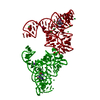
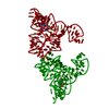
ENGINEERED RESIDUE IN CHAIN A, G 18 TO U ENGINEERED RESIDUE IN CHAIN A, G 19 TO U ENGINEERED ...ENGINEERED RESIDUE IN CHAIN A, G 18 TO U ENGINEERED RESIDUE IN CHAIN A, G 19 TO U ENGINEERED RESIDUE IN CHAIN A, U 34 TO C ENGINEERED RESIDUE IN CHAIN A, A 94 TO G
Sequence detailsMUTATIONS AT RESIDUES 18, 19, 34 AND 94. SEE REMARK 400

-
Experimental details

-
Experiment

ExperimentMethod: X-RAY DIFFRACTION / Number of used crystals: 1

-
Sample preparation

CrystalDensity Matthews: 2.31 Å3/Da / Density % sol: 64 % / Description: NONE
Crystal growpH: 7
Details: 40 MM NA-CACODYLATE (PH 7.0), 12 MM SPERMINE-HCL, 12% (V/V) MPD, 80 MM KCL.

-
Data collection

DiffractionMean temperature: 100 K
Diffraction sourceSource: SYNCHROTRON / Site: ESRF / Beamline: ID14-4 / Wavelength: 0.979
DetectorType: ADSC QUANTUM 315r / Detector: CCD / Date: Dec 6, 2010
RadiationProtocol: SINGLE WAVELENGTH / Monochromatic (M) / Laue (L): M / Scattering type: x-ray
Radiation wavelengthWavelength: 0.979 Å / Relative weight: 1
ReflectionResolution: 2.9→50 Å / Num. obs: 6875 / % possible obs: 93.1 % / Observed criterion σ(I): 2 / Redundancy: 6.1 % / Rmerge(I) obs: 0.05 / Net I/σ(I): 40.6
Reflection shellResolution: 2.9→3 Å / Redundancy: 5.6 % / Rmerge(I) obs: 0.42 / Mean I/σ(I) obs: 2.44 / % possible all: 64.5

-
Processing

Software
NameVersionClassification
REFMAC5.5.0109refinement
HKL-2000data reduction
SCALEPACKdata scaling
REFMACphasing
RefinementMethod to determine structure: MOLECULAR REPLACEMENT
Starting model: PDB ENTRY 3GX5
Resolution: 2.9→57.62 Å / Cor.coef. Fo:Fc: 0.925 / Cor.coef. Fo:Fc free: 0.893 / Cross valid method: THROUGHOUT / ESU R Free: 0.573 / Stereochemistry target values: MAXIMUM LIKELIHOOD
Details: HYDROGENS HAVE BEEN ADDED IN THE RIDING POSITIONS. U VALUES REFINED INDIVIDUALLY
RfactorNum. reflection% reflectionSelection details
Rfree0.32145 666 9.8 %RANDOM
Rwork0.26892 ---
obs0.27406 6121 92.52 %-
Solvent computationIon probe radii: 0.8 Å / Shrinkage radii: 0.8 Å / VDW probe radii: 1.4 Å / Solvent model: MASK
Displacement parametersBiso mean: 88.153 Å2
Baniso -1Baniso -2Baniso -3
1-3.89 Å20 Å20 Å2
2--3.89 Å20 Å2
3----7.78 Å2
Refinement stepCycle: LAST / Resolution: 2.9→57.62 Å
ProteinNucleic acidLigandSolventTotal
Num. atoms0 2024 32 2 2058
Refine LS restraints
Refine-IDTypeDev idealDev ideal targetNumber
X-RAY DIFFRACTIONr_bond_refined_d0.010.0212287
X-RAY DIFFRACTIONr_bond_other_d00.02785
X-RAY DIFFRACTIONr_angle_refined_deg1.91433564
X-RAY DIFFRACTIONr_angle_other_deg1.53132047
X-RAY DIFFRACTIONr_dihedral_angle_1_deg
X-RAY DIFFRACTIONr_dihedral_angle_2_deg
X-RAY DIFFRACTIONr_dihedral_angle_3_deg
X-RAY DIFFRACTIONr_dihedral_angle_4_deg
X-RAY DIFFRACTIONr_chiral_restr0.0740.2475
X-RAY DIFFRACTIONr_gen_planes_refined0.0070.02996
X-RAY DIFFRACTIONr_gen_planes_other0.010.02202
X-RAY DIFFRACTIONr_nbd_refined
X-RAY DIFFRACTIONr_nbd_other
X-RAY DIFFRACTIONr_nbtor_refined
X-RAY DIFFRACTIONr_nbtor_other
X-RAY DIFFRACTIONr_xyhbond_nbd_refined
X-RAY DIFFRACTIONr_xyhbond_nbd_other
X-RAY DIFFRACTIONr_metal_ion_refined
X-RAY DIFFRACTIONr_metal_ion_other
X-RAY DIFFRACTIONr_symmetry_vdw_refined
X-RAY DIFFRACTIONr_symmetry_vdw_other
X-RAY DIFFRACTIONr_symmetry_hbond_refined
X-RAY DIFFRACTIONr_symmetry_hbond_other
X-RAY DIFFRACTIONr_symmetry_metal_ion_refined
X-RAY DIFFRACTIONr_symmetry_metal_ion_other
X-RAY DIFFRACTIONr_mcbond_it
X-RAY DIFFRACTIONr_mcbond_other
X-RAY DIFFRACTIONr_mcangle_it
X-RAY DIFFRACTIONr_mcangle_other
X-RAY DIFFRACTIONr_scbond_it
X-RAY DIFFRACTIONr_scbond_other
X-RAY DIFFRACTIONr_scangle_it
X-RAY DIFFRACTIONr_scangle_other
X-RAY DIFFRACTIONr_long_range_B_refined
X-RAY DIFFRACTIONr_long_range_B_other
X-RAY DIFFRACTIONr_rigid_bond_restr
X-RAY DIFFRACTIONr_sphericity_free
X-RAY DIFFRACTIONr_sphericity_bonded
LS refinement shellResolution: 2.9→2.975 Å / Total num. of bins used: 20
RfactorNum. reflection% reflection
Rfree0.536 28 -
Rwork0.439 304 -
obs--62.29 %

+
About Yorodumi

-
News

-
Feb 9, 2022. New format data for meta-information of EMDB entries

New format data for meta-information of EMDB entries

  • Version 3 of the EMDB header file is now the official format.
  • The previous official version 1.9 will be removed from the archive.

Related info.:EMDB header

External links:wwPDB to switch to version 3 of the EMDB data model

-
Aug 12, 2020. Covid-19 info

Covid-19 info

URL: https://pdbj.org/emnavi/covid19.php

New page: Covid-19 featured information page in EM Navigator.

Related info.:Covid-19 info / Mar 5, 2020. Novel coronavirus structure data

+
Mar 5, 2020. Novel coronavirus structure data

Novel coronavirus structure data

Related info.:Yorodumi Speices / Aug 12, 2020. Covid-19 info

External links:COVID-19 featured content - PDBj / Molecule of the Month (242):Coronavirus Proteases

+
Jan 31, 2019. EMDB accession codes are about to change! (news from PDBe EMDB page)

EMDB accession codes are about to change! (news from PDBe EMDB page)

  • The allocation of 4 digits for EMDB accession codes will soon come to an end. Whilst these codes will remain in use, new EMDB accession codes will include an additional digit and will expand incrementally as the available range of codes is exhausted. The current 4-digit format prefixed with “EMD-” (i.e. EMD-XXXX) will advance to a 5-digit format (i.e. EMD-XXXXX), and so on. It is currently estimated that the 4-digit codes will be depleted around Spring 2019, at which point the 5-digit format will come into force.
  • The EM Navigator/Yorodumi systems omit the EMD- prefix.

Related info.:Q: What is EMD? / ID/Accession-code notation in Yorodumi/EM Navigator

External links:EMDB Accession Codes are Changing Soon! / Contact to PDBj

+
Jul 12, 2017. Major update of PDB

Major update of PDB

  • wwPDB released updated PDB data conforming to the new PDBx/mmCIF dictionary.
  • This is a major update changing the version number from 4 to 5, and with Remediation, in which all the entries are updated.
  • In this update, many items about electron microscopy experimental information are reorganized (e.g. em_software).
  • Now, EM Navigator and Yorodumi are based on the updated data.

External links:wwPDB Remediation / Enriched Model Files Conforming to OneDep Data Standards Now Available in the PDB FTP Archive

-
Yorodumi

Thousand views of thousand structures

  • Yorodumi is a browser for structure data from EMDB, PDB, SASBDB, etc.
  • This page is also the successor to EM Navigator detail page, and also detail information page/front-end page for Omokage search.
  • The word "yorodu" (or yorozu) is an old Japanese word meaning "ten thousand". "mi" (miru) is to see.

Related info.:EMDB / PDB / SASBDB / Comparison of 3 databanks / Yorodumi Search / Aug 31, 2016. New EM Navigator & Yorodumi / Yorodumi Papers / Jmol/JSmol / Function and homology information / Changes in new EM Navigator and Yorodumi

Read more