 Movie
Movie Controller

[English] 日本語
 Yorodumi
- PDB-3gmu: Crystal Structure of Beta-Lactamse Inhibitory Protein (BLIP) in A... -
+ Open data

- Basic information
| Entry | Database: PDB / ID: 3gmu | ||||||
|---|---|---|---|---|---|---|---|
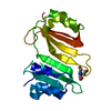
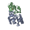
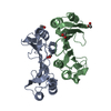
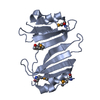
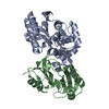
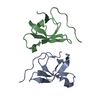
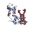
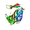
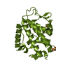
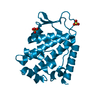
| Title | Crystal Structure of Beta-Lactamse Inhibitory Protein (BLIP) in Apo Form | ||||||
 Components Components | Beta-lactamase inhibitory protein | ||||||
 Keywords Keywords | PROTEIN BINDING / 2-layer alpha/beta sandwich / Disulfide bond / Secreted | ||||||
| Function / homology |  Function and homology information Function and homology information | ||||||
| Biological species |  Streptomyces clavuligerus (bacteria) Streptomyces clavuligerus (bacteria) | ||||||
| Method |  X-RAY DIFFRACTION / MOLECULAR REPLACEMENT / molecular replacement / Resolution: 1.98 Å X-RAY DIFFRACTION / MOLECULAR REPLACEMENT / molecular replacement / Resolution: 1.98 Å | ||||||
 Authors Authors | Strynadka, N.C.J. / Gretes, M. / James, M.N.G. | ||||||
 Citation Citation | Journal: J.Mol.Biol. / Year: 2009 Title: Insights into positive and negative requirements for protein-protein interactions by crystallographic analysis of the beta-lactamase inhibitory proteins BLIP, BLIP-I, and BLP. Authors: Gretes, M. / Lim, D.C. / de Castro, L. / Jensen, S.E. / Kang, S.G. / Lee, K.J. / Strynadka, N.C. #1: Journal: Nature / Year: 1994 Title: Structural and kinetic characterization of a beta-lactamase-inhibitor protein. Authors: Strynadka, N.C. / Jensen, S.E. / Johns, K. / Blanchard, H. / Page, M. / Matagne, A. / Frere, J.M. / James, M.N. | ||||||
| History |
|
- Structure visualization
| Structure viewer | Molecule: MolmilJmol/JSmol |
|---|
- Downloads & links
-Download
| PDBx/mmCIF format | 3gmu.cif.gz | 46.6 KB | Display | PDBx/mmCIF format |
|---|---|---|---|---|
| PDB format | pdb3gmu.ent.gz | 32.4 KB | Display | PDB format |
| PDBx/mmJSON format | 3gmu.json.gz | Tree view | PDBx/mmJSON format | |
| Others |  Other downloads Other downloads |
-Validation report
| Arichive directory | https://data.pdbj.org/pub/pdb/validation_reports/gm/3gmuftp://data.pdbj.org/pub/pdb/validation_reports/gm/3gmu | HTTPS FTP |
|---|
-Related structure data


-Links
 PDBj
PDBj
- Assembly
| Deposited unit | 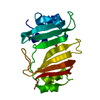
| ||||||||
|---|---|---|---|---|---|---|---|---|---|
| 1 |
| ||||||||
| Unit cell |
| ||||||||
| Components on special symmetry positions |
|
-Components
| #1: Protein | Mass: 17556.492 Da / Num. of mol.: 1 / Source method: isolated from a natural source / Details: cell culture filtrate / Source: (natural) Streptomyces clavuligerus (bacteria) / Strain: NRRL 3585 / References: UniProt: P35804 |
|---|---|
| #2: Chemical | ChemComp-NH4 /   Mass: 18.038 Da / Num. of mol.: 1 / Source method: obtained synthetically / Formula: H4N Mass: 18.038 Da / Num. of mol.: 1 / Source method: obtained synthetically / Formula: H4N |
| #3: Chemical | ChemComp-SO4 /   Mass: 96.063 Da / Num. of mol.: 1 / Source method: obtained synthetically / Formula: SO4 Mass: 96.063 Da / Num. of mol.: 1 / Source method: obtained synthetically / Formula: SO4 |
| #4: Water | ChemComp-HOH /  Mass: 18.015 Da / Num. of mol.: 114 / Source method: isolated from a natural source / Formula: H2O Mass: 18.015 Da / Num. of mol.: 114 / Source method: isolated from a natural source / Formula: H2O |
| Has protein modification | Y |
-Experimental details
-Experiment
| Experiment | Method: X-RAY DIFFRACTION / Number of used crystals: 1 |
|---|
- Sample preparation
| Crystal | Density Matthews: 2.13 Å3/Da / Density % sol: 42.15 % |
|---|---|
| Crystal grow | Temperature: 298 K / Method: vapor diffusion / pH: 5.5 Details: 10 mg/ml protein, 20% saturated ammonium sulfate, 50 mM sodium citrate, pH 5.5, vapor diffusion, temperature 298K |
-Data collection
| Diffraction | Mean temperature: 100 K |
|---|---|
| Diffraction source | Source: ROTATING ANODE / Type: RIGAKU RU200 / Wavelength: 1.54056 Å |
| Detector | Type: SDMS TWIN AREA DETECTOR SYSTEM / Detector: AREA DETECTOR / Date: Dec 17, 1992 |
| Radiation | Monochromator: GRAPHITE / Protocol: SINGLE WAVELENGTH / Scattering type: x-ray |
| Radiation wavelength | Wavelength: 1.54056 Å / Relative weight: 1 |
| Reflection | Resolution: 1.98→59.34 Å / Num. obs: 9038 / % possible obs: 84.6 % / Redundancy: 1.9 % / Rsym value: 0.178 / Net I/σ(I): 7.4 |
| Reflection shell | Resolution: 1.98→2.03 Å / Mean I/σ(I) obs: 1.8 / % possible all: 64.8 |
-Phasing
| Phasing | Method: molecular replacement | |||||||||
|---|---|---|---|---|---|---|---|---|---|---|
| Phasing MR | Model details: Phaser MODE: MR_AUTO
|
- Processing
| Software |
| ||||||||||||||||||||||||||||||||||||||||||||||||||||||||||||||||||||||||||||||||||||||||||
|---|---|---|---|---|---|---|---|---|---|---|---|---|---|---|---|---|---|---|---|---|---|---|---|---|---|---|---|---|---|---|---|---|---|---|---|---|---|---|---|---|---|---|---|---|---|---|---|---|---|---|---|---|---|---|---|---|---|---|---|---|---|---|---|---|---|---|---|---|---|---|---|---|---|---|---|---|---|---|---|---|---|---|---|---|---|---|---|---|---|---|---|
| Refinement | Method to determine structure: MOLECULAR REPLACEMENT / Resolution: 1.98→59.3 Å / Cor.coef. Fo:Fc: 0.953 / Cor.coef. Fo:Fc free: 0.911 / WRfactor Rfree: 0.221 / WRfactor Rwork: 0.149 / Occupancy max: 1 / Occupancy min: 0.5 / FOM work R set: 0.863 / SU B: 3.915 / SU ML: 0.115 / SU R Cruickshank DPI: 0.228 / SU Rfree: 0.193 / Cross valid method: THROUGHOUT / σ(F): 0 / ESU R: 0.228 / ESU R Free: 0.193 / Stereochemistry target values: MAXIMUM LIKELIHOOD / Details: HYDROGENS HAVE BEEN ADDED IN THE RIDING POSITIONS
| ||||||||||||||||||||||||||||||||||||||||||||||||||||||||||||||||||||||||||||||||||||||||||
| Solvent computation | Ion probe radii: 0.8 Å / Shrinkage radii: 0.8 Å / VDW probe radii: 1.2 Å / Solvent model: MASK | ||||||||||||||||||||||||||||||||||||||||||||||||||||||||||||||||||||||||||||||||||||||||||
| Displacement parameters | Biso max: 56.93 Å2 / Biso mean: 18.946 Å2 / Biso min: 2 Å2
| ||||||||||||||||||||||||||||||||||||||||||||||||||||||||||||||||||||||||||||||||||||||||||
| Refinement step | Cycle: LAST / Resolution: 1.98→59.3 Å
| ||||||||||||||||||||||||||||||||||||||||||||||||||||||||||||||||||||||||||||||||||||||||||
| Refine LS restraints |
| ||||||||||||||||||||||||||||||||||||||||||||||||||||||||||||||||||||||||||||||||||||||||||
| LS refinement shell | Resolution: 1.98→2.03 Å / Total num. of bins used: 20
|
