 Movie
Movie Controller

[English] 日本語
 Yorodumi
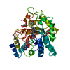
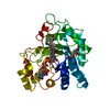
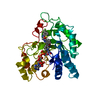
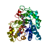
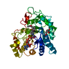
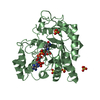
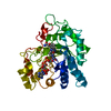
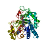
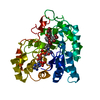
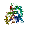
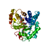
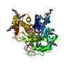
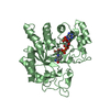
- PDB-3ghu: Human aldose reductase in complex with NADP+ and the inhibitor ID... -
+ Open data

- Basic information
| Entry | Database: PDB / ID: 3ghu | ||||||
|---|---|---|---|---|---|---|---|
| Title | Human aldose reductase in complex with NADP+ and the inhibitor IDD594. Investigation of global effects of radiation damage on protein structure. Forth stage of radiation damage. | ||||||
 Components Components | Aldose reductase | ||||||
 Keywords Keywords | OXIDOREDUCTASE / Acetylation / Cataract / Cytoplasm / NADP / Phosphoprotein / Polymorphism | ||||||
| Function / homology |  Function and homology information Function and homology informationglyceraldehyde oxidoreductase activity / Fructose biosynthesis / fructose biosynthetic process / L-glucuronate reductase activity / aldose reductase / D/L-glyceraldehyde reductase / glycerol dehydrogenase (NADP+) activity / C21-steroid hormone biosynthetic process / NADP-retinol dehydrogenase / Pregnenolone biosynthesis ...glyceraldehyde oxidoreductase activity / Fructose biosynthesis / fructose biosynthetic process / L-glucuronate reductase activity / aldose reductase / D/L-glyceraldehyde reductase / glycerol dehydrogenase (NADP+) activity / C21-steroid hormone biosynthetic process / NADP-retinol dehydrogenase / Pregnenolone biosynthesis / allyl-alcohol dehydrogenase / allyl-alcohol dehydrogenase activity / Galactose catabolism / prostaglandin H2 endoperoxidase reductase activity / regulation of urine volume / all-trans-retinol dehydrogenase (NADP+) activity / metanephric collecting duct development / daunorubicin metabolic process / doxorubicin metabolic process / retinal dehydrogenase (NAD+) activity / epithelial cell maturation / aldose reductase (NADPH) activity / cellular hyperosmotic salinity response / renal water homeostasis / retinoid metabolic process / carbohydrate metabolic process / electron transfer activity / negative regulation of apoptotic process / mitochondrion / : / extracellular exosome / nucleoplasm / cytosol Similarity search - Function | ||||||
| Biological species |  Homo sapiens (human) Homo sapiens (human) | ||||||
| Method |  X-RAY DIFFRACTION / SYNCHROTRON / MOLECULAR REPLACEMENT / Resolution: 1.2 Å X-RAY DIFFRACTION / SYNCHROTRON / MOLECULAR REPLACEMENT / Resolution: 1.2 Å | ||||||
 Authors Authors | Petrova, T. / Ginell, S. / Hazemann, I. / Mitschler, A. / Podjarny, A. / Joachimiak, A. | ||||||
 Citation Citation | Journal: J.Mol.Biol. / Year: 2009 Title: X-ray-radiation-induced cooperative atomic movements in protein. Authors: Petrova, T. / Lunin, V.Y. / Ginell, S. / Hazemann, I. / Lazarski, K. / Mitschler, A. / Podjarny, A. / Joachimiak, A. | ||||||
| History |
|
- Structure visualization
| Structure viewer | Molecule: MolmilJmol/JSmol |
|---|
- Downloads & links
-Download
| PDBx/mmCIF format | 3ghu.cif.gz | 197.9 KB | Display | PDBx/mmCIF format |
|---|---|---|---|---|
| PDB format | pdb3ghu.ent.gz | 157.6 KB | Display | PDB format |
| PDBx/mmJSON format | 3ghu.json.gz | Tree view | PDBx/mmJSON format | |
| Others |  Other downloads Other downloads |
-Validation report
| Arichive directory | https://data.pdbj.org/pub/pdb/validation_reports/gh/3ghuftp://data.pdbj.org/pub/pdb/validation_reports/gh/3ghu | HTTPS FTP |
|---|
-Related structure data
| Related structure data | 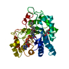 3ghrC  3ghsC 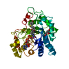 3ghtC  1us0S S: Starting model for refinement C: citing same article ( |
|---|---|
| Similar structure data |


-Links
 PDBj
PDBj


- Assembly
| Deposited unit | 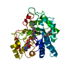
| ||||||||
|---|---|---|---|---|---|---|---|---|---|
| 1 |
| ||||||||
| Unit cell |
|
-Components
| #1: Protein | Mass: 35898.340 Da / Num. of mol.: 1 Source method: isolated from a genetically manipulated source Source: (gene. exp.) Homo sapiens (human) / Gene: AKR1B1, ALDR1 / Production host:  |
|---|---|
| #2: Chemical | ChemComp-NDP /   Mass: 745.421 Da / Num. of mol.: 1 / Source method: obtained synthetically / Formula: C21H30N7O17P3 Mass: 745.421 Da / Num. of mol.: 1 / Source method: obtained synthetically / Formula: C21H30N7O17P3 |
| #3: Chemical | ChemComp-LDT / 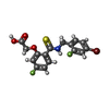  Mass: 416.237 Da / Num. of mol.: 1 / Source method: obtained synthetically / Formula: C16H12BrF2NO3S Mass: 416.237 Da / Num. of mol.: 1 / Source method: obtained synthetically / Formula: C16H12BrF2NO3S |
| #4: Chemical | ChemComp-CIT /   Mass: 192.124 Da / Num. of mol.: 1 / Source method: obtained synthetically / Formula: C6H8O7 Mass: 192.124 Da / Num. of mol.: 1 / Source method: obtained synthetically / Formula: C6H8O7 |
| #5: Water | ChemComp-HOH /  Mass: 18.015 Da / Num. of mol.: 630 / Source method: isolated from a natural source / Formula: H2O Mass: 18.015 Da / Num. of mol.: 630 / Source method: isolated from a natural source / Formula: H2O |
| Sequence details | AUTHORS STATE THAT RESIDUE NUMBER 4 IS ILE. ELECTRON DENSITY FOR THIS RESIDUE IS VERY CLEAR |
-Experimental details
-Experiment
| Experiment | Method: X-RAY DIFFRACTION / Number of used crystals: 1 |
|---|
- Sample preparation
| Crystal | Density Matthews: 2.18 Å3/Da / Density % sol: 43.65 % |
|---|---|
| Crystal grow | Temperature: 277 K / Method: vapor diffusion, hanging drop / pH: 5 Details: The co-crystallization with IDD594 was carried out at room temperature (ratios protein/coenzyme/inhibitor = 1/2/2), pH 5.0, VAPOR DIFFUSION, HANGING DROP, temperature 277K |
-Data collection
| Diffraction | Mean temperature: 100 K |
|---|---|
| Diffraction source | Source: SYNCHROTRON / Site: APS  / Beamline: 19-ID / Wavelength: 0.91996 Å / Beamline: 19-ID / Wavelength: 0.91996 Å |
| Detector | Type: ADSC QUANTUM Q315r / Detector: CCD / Date: Aug 20, 2007 Details: 1.02-M FLAT MIRROR MADE OF ZERODUR PROVIDING VERTICAL FOCUSING AND REJECTION OF HARMONIC CONTAMINATION |
| Radiation | Monochromator: DOUBLE CRYSTAL MONOCHROMATOR UTILIZING A SI-111 AND SAGITAL HORIZONTAL FOCUSING Protocol: SINGLE WAVELENGTH / Monochromatic (M) / Laue (L): M / Scattering type: x-ray |
| Radiation wavelength | Wavelength: 0.91996 Å / Relative weight: 1 |
| Reflection | Resolution: 1.2→50 Å / Num. all: 92143 / Num. obs: 92143 / % possible obs: 96.5 % / Observed criterion σ(F): 0 / Observed criterion σ(I): 0 / Redundancy: 3.7 % / Rmerge(I) obs: 0.023 / Rsym value: 0.033 / Net I/σ(I): 33.8 |
| Reflection shell | Resolution: 1.2→1.23 Å / Redundancy: 3.4 % / Rmerge(I) obs: 0.345 / Mean I/σ(I) obs: 3 / Num. unique all: 5869 / % possible all: 93.1 |
- Processing
| Software |
| |||||||||||||||||||||||||
|---|---|---|---|---|---|---|---|---|---|---|---|---|---|---|---|---|---|---|---|---|---|---|---|---|---|---|
| Refinement | Method to determine structure: MOLECULAR REPLACEMENT Starting model: 1US0 Resolution: 1.2→50 Å / σ(F): 4 / σ(I): 0 / Stereochemistry target values: Engh & Huber
| |||||||||||||||||||||||||
| Refinement step | Cycle: LAST / Resolution: 1.2→50 Å
|
