Mass: 18.015 Da / Num. of mol.: 431 / Source method: isolated from a natural source / Formula: H2O
-
Experimental details
-
Experiment
Experiment
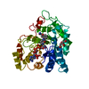
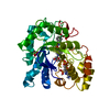
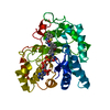
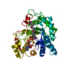
Method: X-RAY DIFFRACTION / Number of used crystals: 1
-
Sample preparation
Crystal
Density Matthews: 2.18 Å3/Da / Density % sol: 43.49 %
Crystal grow
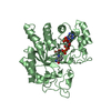
Temperature: 298 K / pH: 4.2 Details: The hanging drop method was used to obtain crystals (EasyXtal Tool 24 culture plates, Qiagen). WT hALR2 was co-crystallized with the oxidized form of coenzyme NADP+ (Sigma) and inhibitor 2 ...Details: The hanging drop method was used to obtain crystals (EasyXtal Tool 24 culture plates, Qiagen). WT hALR2 was co-crystallized with the oxidized form of coenzyme NADP+ (Sigma) and inhibitor 2 at room temperature (ratios of protein / inhibitor / coenzyme = 1/2/2). Hanging drops were made by the mixing mother liquor (0.06 M citric acid, 0.04 M Bis-tris propane, 12% (w/v) PEG 3350, pH 4.2) with holoenzyme solution (10 mg/mL, 20 mM hepes, pH 7.0). Crystals were observed after three days of equilibration, EVAPORATION, temperature 298K
In the structure databanks used in Yorodumi, some data are registered as the other names, "COVID-19 virus" and "2019-nCoV". Here are the details of the virus and the list of structure data.
Jan 31, 2019. EMDB accession codes are about to change! (news from PDBe EMDB page)
EMDB accession codes are about to change! (news from PDBe EMDB page)
The allocation of 4 digits for EMDB accession codes will soon come to an end. Whilst these codes will remain in use, new EMDB accession codes will include an additional digit and will expand incrementally as the available range of codes is exhausted. The current 4-digit format prefixed with “EMD-” (i.e. EMD-XXXX) will advance to a 5-digit format (i.e. EMD-XXXXX), and so on. It is currently estimated that the 4-digit codes will be depleted around Spring 2019, at which point the 5-digit format will come into force.
The EM Navigator/Yorodumi systems omit the EMD- prefix.
Related info.:Q: What is EMD? / ID/Accession-code notation in Yorodumi/EM Navigator
Yorodumi is a browser for structure data from EMDB, PDB, SASBDB, etc.
This page is also the successor to EM Navigator detail page, and also detail information page/front-end page for Omokage search.
The word "yorodu" (or yorozu) is an old Japanese word meaning "ten thousand". "mi" (miru) is to see.
Related info.:EMDB / PDB / SASBDB / Comparison of 3 databanks / Yorodumi Search / Aug 31, 2016. New EM Navigator & Yorodumi / Yorodumi Papers / Jmol/JSmol / Function and homology information / Changes in new EM Navigator and Yorodumi
 Movie
Movie Controller

 Yorodumi
 Open data
 Basic information
 Components
 Keywords
 Function and homology information
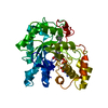
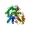
 Homo sapiens (human)
 X-RAY DIFFRACTION /
  Authors
 Citation
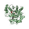
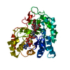
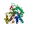
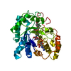
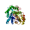
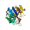
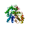
 Structure visualization
 Downloads & links
 Other downloads
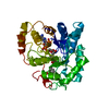


 PDBj
PDBj
 Assembly


 Mass: 743.405 Da / Num. of mol.: 1 / Source method: obtained synthetically / Formula: C21H28N7O17P3
 Mass: 389.747 Da / Num. of mol.: 1 / Source method: obtained synthetically / Formula: C17H12ClN3O6
 Mass: 192.124 Da / Num. of mol.: 1 / Source method: obtained synthetically / Formula: C6H8O7
 Mass: 18.015 Da / Num. of mol.: 431 / Source method: isolated from a natural source / Formula: H2O
 Sample preparation
 / Beamline: BL9-2 / Wavelength: 0.979
  Processing