 Movie
Movie Controller

[English] 日本語
 Yorodumi
- PDB-3gc6: Structural insights into the catalytic mechanism of CD38: Evidenc... -
+ Open data

- Basic information
| Entry | Database: PDB / ID: 3gc6 | |||||||||
|---|---|---|---|---|---|---|---|---|---|---|
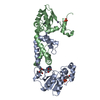
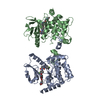
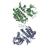
| Title | Structural insights into the catalytic mechanism of CD38: Evidence for a conformationally flexible covalent enzyme-substrate complex. | |||||||||
 Components Components | Ecto-NAD+ glycohydrolase (CD38 molecule) | |||||||||
 Keywords Keywords | HYDROLASE / CD38 / cyclic ADP ribose / ecto-ADP-ribosyl cyclase / Glycosidase | |||||||||
| Function / homology |  Function and homology information Function and homology information2'-phospho-ADP-ribosyl cyclase/2'-phospho-cyclic-ADP-ribose transferase / ADP-ribosyl cyclase/cyclic ADP-ribose hydrolase / NAD+ nucleosidase activity, cyclic ADP-ribose generating / transferase activity / membrane Similarity search - Function | |||||||||
| Biological species |  | |||||||||
| Method |  X-RAY DIFFRACTION / SYNCHROTRON / MOLECULAR REPLACEMENT / Resolution: 1.51 Å X-RAY DIFFRACTION / SYNCHROTRON / MOLECULAR REPLACEMENT / Resolution: 1.51 Å | |||||||||
 Authors Authors | Egea, P.F. / Muller-Steffner, H. / Stroud, R.M. / Oppenheimer, N. / Kellenberger, E. / Schuber, F. | |||||||||
 Citation Citation | Journal: Plos One / Year: 2012 Title: Insights into the mechanism of bovine CD38/NAD+glycohydrolase from the X-ray structures of its Michaelis complex and covalently-trapped intermediates. Authors: Egea, P.F. / Muller-Steffner, H. / Kuhn, I. / Cakir-Kiefer, C. / Oppenheimer, N.J. / Stroud, R.M. / Kellenberger, E. / Schuber, F. | |||||||||
| History |
|
- Structure visualization
| Structure viewer | Molecule: MolmilJmol/JSmol |
|---|
- Downloads & links
-Download
| PDBx/mmCIF format | 3gc6.cif.gz | 175.2 KB | Display | PDBx/mmCIF format |
|---|---|---|---|---|
| PDB format | pdb3gc6.ent.gz | 128.7 KB | Display | PDB format |
| PDBx/mmJSON format | 3gc6.json.gz | Tree view | PDBx/mmJSON format | |
| Others |  Other downloads Other downloads |
-Validation report
| Arichive directory | https://data.pdbj.org/pub/pdb/validation_reports/gc/3gc6ftp://data.pdbj.org/pub/pdb/validation_reports/gc/3gc6 | HTTPS FTP |
|---|
-Related structure data
| Related structure data |  3gh3C  3ghhC  3kouC  3p5sC 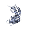 1yh3S S: Starting model for refinement C: citing same article ( |
|---|---|
| Similar structure data |


-Links
 PDBj
PDBj
- Assembly
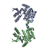
| Deposited unit | 
| ||||||||
|---|---|---|---|---|---|---|---|---|---|
| 1 | 
| ||||||||
| 2 |
| ||||||||
| Unit cell |
|
-Components
| #1: Protein | Mass: 28300.061 Da / Num. of mol.: 2 / Mutation: E218Q Source method: isolated from a genetically manipulated source Source: (gene. exp.)  Pichia pastoris (fungus) / References: UniProt: Q9TTF5, NAD+ glycohydrolase Pichia pastoris (fungus) / References: UniProt: Q9TTF5, NAD+ glycohydrolase#2: Polysaccharide | Source method: isolated from a genetically manipulated source #3: Chemical | ChemComp-SO4 /   Mass: 96.063 Da / Num. of mol.: 5 / Source method: obtained synthetically / Formula: SO4 Mass: 96.063 Da / Num. of mol.: 5 / Source method: obtained synthetically / Formula: SO4#4: Water | ChemComp-HOH / |  Mass: 18.015 Da / Num. of mol.: 488 / Source method: isolated from a natural source / Formula: H2O Mass: 18.015 Da / Num. of mol.: 488 / Source method: isolated from a natural source / Formula: H2OHas protein modification | Y | |
|---|
-Experimental details
-Experiment
| Experiment | Method: X-RAY DIFFRACTION / Number of used crystals: 1 |
|---|
- Sample preparation
| Crystal | Density Matthews: 2.54 Å3/Da / Density % sol: 51.58 % |
|---|---|
| Crystal grow | Temperature: 293 K / Method: vapor diffusion, hanging drop / pH: 6 Details: 20-30% PEG 4000, 50-250mM Ammonium Sulfate, 100 mM sodium cacodylate or sodium acetate or MES at pH-6.0-6.5, VAPOR DIFFUSION, HANGING DROP, temperature 293K |
-Data collection
| Diffraction | Mean temperature: 77 K |
|---|---|
| Diffraction source | Source: SYNCHROTRON / Site: ALS  / Beamline: 8.3.1 / Beamline: 8.3.1 |
| Detector | Type: ADSC QUANTUM 315 / Detector: CCD / Date: Apr 25, 2007 |
| Radiation | Protocol: 1.11587 / Monochromatic (M) / Laue (L): M / Scattering type: x-ray |
| Radiation wavelength | Relative weight: 1 |
| Reflection | Resolution: 1.51→80.1 Å / Num. all: 74734 / Num. obs: 74734 / % possible obs: 82 % / Redundancy: 3.3 % / Biso Wilson estimate: 21 Å2 / Rsym value: 0.035 / Net I/σ(I): 16.9 |
| Reflection shell | Resolution: 1.51→1.61 Å / Redundancy: 1.3 % / Mean I/σ(I) obs: 1.5 / Num. unique all: 4271 / Rsym value: 0.318 / % possible all: 33 |
- Processing
| Software |
| |||||||||||||||||||||||||||||||||||||||||||||||||||||||||||||||||||||||||||
|---|---|---|---|---|---|---|---|---|---|---|---|---|---|---|---|---|---|---|---|---|---|---|---|---|---|---|---|---|---|---|---|---|---|---|---|---|---|---|---|---|---|---|---|---|---|---|---|---|---|---|---|---|---|---|---|---|---|---|---|---|---|---|---|---|---|---|---|---|---|---|---|---|---|---|---|---|
| Refinement | Method to determine structure: MOLECULAR REPLACEMENT Starting model: PDB entry 1YH3 Resolution: 1.51→44.95 Å / Cor.coef. Fo:Fc: 0.959 / Cor.coef. Fo:Fc free: 0.952 / SU B: 2.541 / SU ML: 0.047 / Cross valid method: THROUGHOUT / ESU R: 0.094 / ESU R Free: 0.09 / Stereochemistry target values: MAXIMUM LIKELIHOOD / Details: HYDROGENS HAVE BEEN ADDED IN THE RIDING POSITIONS
| |||||||||||||||||||||||||||||||||||||||||||||||||||||||||||||||||||||||||||
| Solvent computation | Ion probe radii: 0.8 Å / Shrinkage radii: 0.8 Å / VDW probe radii: 1.2 Å / Solvent model: MASK | |||||||||||||||||||||||||||||||||||||||||||||||||||||||||||||||||||||||||||
| Displacement parameters | Biso mean: 22.542 Å2
| |||||||||||||||||||||||||||||||||||||||||||||||||||||||||||||||||||||||||||
| Refinement step | Cycle: LAST / Resolution: 1.51→44.95 Å
| |||||||||||||||||||||||||||||||||||||||||||||||||||||||||||||||||||||||||||
| Refine LS restraints |
| |||||||||||||||||||||||||||||||||||||||||||||||||||||||||||||||||||||||||||
| LS refinement shell | Resolution: 1.51→1.549 Å / Total num. of bins used: 20
| |||||||||||||||||||||||||||||||||||||||||||||||||||||||||||||||||||||||||||
| Refinement TLS params. | Method: refined / Refine-ID: X-RAY DIFFRACTION
| |||||||||||||||||||||||||||||||||||||||||||||||||||||||||||||||||||||||||||
| Refinement TLS group |
|
