| Entry | Database: PDB / ID: 5owq
|
|---|
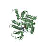
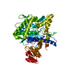
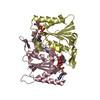
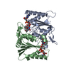
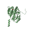
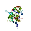
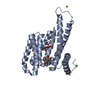
| Title | Human STK10 bound to dovitinib |
|---|
 Components Components | Serine/threonine-protein kinase 10 |
|---|
 Keywords Keywords | TRANSFERASE / Kinase / Structural Genomics Consortium / SGC |
|---|
| Function / homology |  Function and homology information Function and homology information
lymphocyte aggregation / regulation of lymphocyte migration / RHOB GTPase cycle / RHOC GTPase cycle / RHOA GTPase cycle / specific granule membrane / protein autophosphorylation / protein phosphorylation / non-specific serine/threonine protein kinase / intracellular signal transduction ...lymphocyte aggregation / regulation of lymphocyte migration / RHOB GTPase cycle / RHOC GTPase cycle / RHOA GTPase cycle / specific granule membrane / protein autophosphorylation / protein phosphorylation / non-specific serine/threonine protein kinase / intracellular signal transduction / nuclear body / protein serine kinase activity / protein serine/threonine kinase activity / Neutrophil degranulation / protein homodimerization activity / extracellular exosome / nucleoplasm / ATP binding / identical protein binding / plasma membrane / cytoplasm / cytosolSimilarity search - Function Serine/threonine-protein kinase 10, catalytic domain / Polo kinase kinase / : / Polo kinase kinase / Phosphorylase Kinase; domain 1 / Phosphorylase Kinase; domain 1 / Transferase(Phosphotransferase) domain 1 / Transferase(Phosphotransferase); domain 1 / Serine/threonine-protein kinase, active site / Serine/Threonine protein kinases active-site signature. ...Serine/threonine-protein kinase 10, catalytic domain / Polo kinase kinase / : / Polo kinase kinase / Phosphorylase Kinase; domain 1 / Phosphorylase Kinase; domain 1 / Transferase(Phosphotransferase) domain 1 / Transferase(Phosphotransferase); domain 1 / Serine/threonine-protein kinase, active site / Serine/Threonine protein kinases active-site signature. / Protein kinase domain / Serine/Threonine protein kinases, catalytic domain / Protein kinase, ATP binding site / Protein kinases ATP-binding region signature. / Protein kinase domain profile. / Protein kinase domain / Protein kinase-like domain superfamily / 2-Layer Sandwich / Orthogonal Bundle / Mainly Alpha / Alpha BetaSimilarity search - Domain/homology |
|---|
| Biological species |  Homo sapiens (human) Homo sapiens (human) |
|---|
| Method |  X-RAY DIFFRACTION / SYNCHROTRON / MOLECULAR REPLACEMENT / Resolution: 2.7 Å X-RAY DIFFRACTION / SYNCHROTRON / MOLECULAR REPLACEMENT / Resolution: 2.7 Å |
|---|
 Authors Authors | Szklarz, M. / von Delft, F. / Bountra, C. / Knapp, S. / Edwards, A.M. / Arrowsmith, C. / Elkins, J.M. |
|---|
 Citation Citation | Journal: To Be Published
Title: Human STK10 bound to dovitinib
Authors: Elkins, J.M. |
|---|
| History | | Deposition | Sep 4, 2017 | Deposition site: PDBE / Processing site: PDBE |
|---|
| Revision 1.0 | Sep 13, 2017 | Provider: repository / Type: Initial release |
|---|
| Revision 1.1 | Jan 24, 2018 | Group: Source and taxonomy / Category: entity_src_gen / Item: _entity_src_gen.pdbx_host_org_scientific_name |
|---|
| Revision 1.2 | May 1, 2024 | Group: Data collection / Database references / Refinement description
Category: chem_comp_atom / chem_comp_bond ...chem_comp_atom / chem_comp_bond / database_2 / pdbx_initial_refinement_model / struct_ncs_dom_lim
Item: _database_2.pdbx_DOI / _database_2.pdbx_database_accession ..._database_2.pdbx_DOI / _database_2.pdbx_database_accession / _struct_ncs_dom_lim.beg_auth_comp_id / _struct_ncs_dom_lim.beg_label_asym_id / _struct_ncs_dom_lim.beg_label_comp_id / _struct_ncs_dom_lim.beg_label_seq_id / _struct_ncs_dom_lim.end_auth_comp_id / _struct_ncs_dom_lim.end_label_asym_id / _struct_ncs_dom_lim.end_label_comp_id / _struct_ncs_dom_lim.end_label_seq_id |
|---|
| Revision 1.3 | Oct 23, 2024 | Group: Structure summary / Category: pdbx_entry_details / pdbx_modification_feature |
|---|
|
|---|
 Movie
Movie Controller


 Open data
 Basic information
 Structure visualization
 Downloads & links
 Other downloads


 PDBj
PDBj
 Assembly
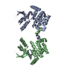


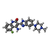
 Mass: 392.429 Da / Num. of mol.: 2 / Source method: obtained synthetically / Formula: C21H21FN6O
 Sample preparation
 / Beamline: I04-1 / Wavelength: 0.9173 Å
 Processing