 Movie
Movie Controller

[English] 日本語
 Yorodumi
- PDB-3p5s: Structural insights into the catalytic mechanism of CD38: Evidenc... -
+ Open data

- Basic information
| Entry | Database: PDB / ID: 3p5s | ||||||
|---|---|---|---|---|---|---|---|
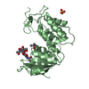
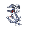
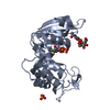
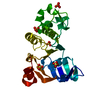
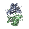
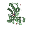
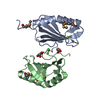
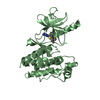
| Title | Structural insights into the catalytic mechanism of CD38: Evidence for a conformationally flexible covalent enzyme-substrate complex | ||||||
 Components Components | CD38 molecule | ||||||
 Keywords Keywords | HYDROLASE / CD38 / CYCLIC ADP RIBOSE / ECTO-ADP-RIBOSYL CYCLASE / GLYCOSIDASE | ||||||
| Function / homology |  Function and homology information Function and homology information2'-phospho-ADP-ribosyl cyclase/2'-phospho-cyclic-ADP-ribose transferase / ADP-ribosyl cyclase/cyclic ADP-ribose hydrolase / NAD+ nucleosidase activity, cyclic ADP-ribose generating / transferase activity / membrane Similarity search - Function | ||||||
| Biological species |  | ||||||
| Method |  X-RAY DIFFRACTION / SYNCHROTRON / MOLECULAR REPLACEMENT / Resolution: 1.95 Å X-RAY DIFFRACTION / SYNCHROTRON / MOLECULAR REPLACEMENT / Resolution: 1.95 Å | ||||||
 Authors Authors | Egea, P.F. / Muller-Stauffler, H. / Kohn, I. / Cakou-Kefir, C. / Stroud, R.M. / Kellenberburger, E. / Schuber, F. | ||||||
 Citation Citation | Journal: Plos One / Year: 2012 Title: Insights into the mechanism of bovine CD38/NAD+glycohydrolase from the X-ray structures of its Michaelis complex and covalently-trapped intermediates. Authors: Egea, P.F. / Muller-Steffner, H. / Kuhn, I. / Cakir-Kiefer, C. / Oppenheimer, N.J. / Stroud, R.M. / Kellenberger, E. / Schuber, F. | ||||||
| History |
|
- Structure visualization
| Structure viewer | Molecule: MolmilJmol/JSmol |
|---|
- Downloads & links
-Download
| PDBx/mmCIF format | 3p5s.cif.gz | 120 KB | Display | PDBx/mmCIF format |
|---|---|---|---|---|
| PDB format | pdb3p5s.ent.gz | 92.4 KB | Display | PDB format |
| PDBx/mmJSON format | 3p5s.json.gz | Tree view | PDBx/mmJSON format | |
| Others |  Other downloads Other downloads |
-Validation report
| Arichive directory | https://data.pdbj.org/pub/pdb/validation_reports/p5/3p5sftp://data.pdbj.org/pub/pdb/validation_reports/p5/3p5s | HTTPS FTP |
|---|
-Related structure data
| Related structure data |  3gc6SC  3gh3C  3ghhC  3kouC C: citing same article ( S: Starting model for refinement |
|---|---|
| Similar structure data |


-Links
 PDBj
PDBj
- Assembly
| Deposited unit | 
| ||||||||
|---|---|---|---|---|---|---|---|---|---|
| 1 | 
| ||||||||
| 2 | 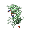
| ||||||||
| Unit cell |
|
-Components
| #1: Protein | Mass: 31587.125 Da / Num. of mol.: 2 Source method: isolated from a genetically manipulated source Source: (gene. exp.)  Pichia pastoris (fungus) / References: UniProt: Q9TTF5, NAD+ glycohydrolase Pichia pastoris (fungus) / References: UniProt: Q9TTF5, NAD+ glycohydrolase#2: Sugar |   Type: D-saccharide, beta linking / Mass: 221.208 Da / Num. of mol.: 2 Type: D-saccharide, beta linking / Mass: 221.208 Da / Num. of mol.: 2Source method: isolated from a genetically manipulated source Formula: C8H15NO6 #3: Chemical |   Mass: 96.063 Da / Num. of mol.: 2 / Source method: obtained synthetically / Formula: SO4 Mass: 96.063 Da / Num. of mol.: 2 / Source method: obtained synthetically / Formula: SO4#4: Chemical |   Mass: 545.307 Da / Num. of mol.: 2 / Source method: obtained synthetically / Formula: C15H22FN5O12P2 Mass: 545.307 Da / Num. of mol.: 2 / Source method: obtained synthetically / Formula: C15H22FN5O12P2#5: Water | ChemComp-HOH / |  Mass: 18.015 Da / Num. of mol.: 341 / Source method: isolated from a natural source / Formula: H2O Mass: 18.015 Da / Num. of mol.: 341 / Source method: isolated from a natural source / Formula: H2OHas protein modification | Y | |
|---|
-Experimental details
-Experiment
| Experiment | Method: X-RAY DIFFRACTION / Number of used crystals: 1 |
|---|
- Sample preparation
| Crystal | Density Matthews: 2.31 Å3/Da / Density % sol: 46.71 % |
|---|---|
| Crystal grow | Temperature: 293 K / Method: vapor diffusion, hanging drop Details: 20-30% PEG 4000, 50-250 mM AMMONIUM SULFATE, 100 MM SODIUM CACODYLATE OR SODIUM ACETATE OR MES AT PH-6.0-6.5, pH 6.0 6.5, VAPOR DIFFUSION, HANGING DROP, temperature 293K PH range: 6.0 6.5 |
-Data collection
| Diffraction | Mean temperature: 130 K |
|---|---|
| Diffraction source | Source: SYNCHROTRON / Site: ALS  / Beamline: 8.3.1 / Wavelength: 1.11587 Å / Beamline: 8.3.1 / Wavelength: 1.11587 Å |
| Detector | Type: ADSC QUANTUM 315r / Detector: CCD / Date: Apr 25, 2007 / Details: could not see them |
| Radiation | Protocol: SINGLE WAVELENGTH / Monochromatic (M) / Laue (L): M / Scattering type: x-ray |
| Radiation wavelength | Wavelength: 1.11587 Å / Relative weight: 1 |
| Reflection | Resolution: 1.95→78.8 Å / Num. all: 110000 / Num. obs: 39086 / % possible obs: 90 % / Observed criterion σ(F): 1 / Observed criterion σ(I): 1 / Redundancy: 2.9 % / Biso Wilson estimate: 26 Å2 / Rsym value: 0.084 / Net I/σ(I): 1 |
- Processing
| Software |
| |||||||||||||||||||||||||||||||||||||||||||||||||||||||||||||||||||||||||||||
|---|---|---|---|---|---|---|---|---|---|---|---|---|---|---|---|---|---|---|---|---|---|---|---|---|---|---|---|---|---|---|---|---|---|---|---|---|---|---|---|---|---|---|---|---|---|---|---|---|---|---|---|---|---|---|---|---|---|---|---|---|---|---|---|---|---|---|---|---|---|---|---|---|---|---|---|---|---|---|
| Refinement | Method to determine structure: MOLECULAR REPLACEMENT Starting model: PDB entry 3GC6 Resolution: 1.95→40.014 Å / SU ML: 0.29 / Cross valid method: random throughout, test set / σ(F): 0 / σ(I): 1 / Stereochemistry target values: ML
| |||||||||||||||||||||||||||||||||||||||||||||||||||||||||||||||||||||||||||||
| Solvent computation | Shrinkage radii: 0.83 Å / VDW probe radii: 1.1 Å / Solvent model: FLAT BULK SOLVENT MODEL / Bsol: 41.574 Å2 / ksol: 0.362 e/Å3 | |||||||||||||||||||||||||||||||||||||||||||||||||||||||||||||||||||||||||||||
| Displacement parameters |
| |||||||||||||||||||||||||||||||||||||||||||||||||||||||||||||||||||||||||||||
| Refinement step | Cycle: LAST / Resolution: 1.95→40.014 Å
| |||||||||||||||||||||||||||||||||||||||||||||||||||||||||||||||||||||||||||||
| Refine LS restraints |
| |||||||||||||||||||||||||||||||||||||||||||||||||||||||||||||||||||||||||||||
| LS refinement shell |
|
