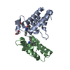
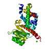
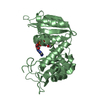
[English] 日本語
Yorodumi
- PDB-4c3z: Nucleotide-free crystal structure of nucleotide-binding domain 1 ... -

+
Open data


ID or keywords:

Loading...

-
Basic information

Entry
Database: PDB / ID: 4c3z
TitleNucleotide-free crystal structure of nucleotide-binding domain 1 from human MRP1 supports a general-base catalysis mechanism for ATP hydrolysis.
ComponentsMULTIDRUG RESISTANCE-ASSOCIATED PROTEIN 1
KeywordsTRANSPORT PROTEIN / ATP-BINDING CASSETTE TRANSPORTERS / MULTIDRUG RESISTANCE PROTEIN 1 / GENERAL-BASE MECHANISM
Function / homology
Function and homology information


sphingolipid translocation / cyclic nucleotide transport / Transport of RCbl within the body / ABC-type vitamin B12 transporter activity / carboxylic acid transmembrane transport / carboxylic acid transmembrane transporter activity / sphingolipid transporter activity / cobalamin transport / leukotriene transport / glutathione transmembrane transport ...sphingolipid translocation / cyclic nucleotide transport / Transport of RCbl within the body / ABC-type vitamin B12 transporter activity / carboxylic acid transmembrane transport / carboxylic acid transmembrane transporter activity / sphingolipid transporter activity / cobalamin transport / leukotriene transport / glutathione transmembrane transport / glutathione transmembrane transporter activity / ABC-type glutathione-S-conjugate transporter / ABC-type glutathione S-conjugate transporter activity / leukotriene metabolic process / ATPase-coupled inorganic anion transmembrane transporter activity / Synthesis of Leukotrienes (LT) and Eoxins (EX) / sphingolipid biosynthetic process / Sphingolipid de novo biosynthesis / heme catabolic process / xenobiotic transport across blood-brain barrier / transepithelial transport / export across plasma membrane / ABC-type xenobiotic transporter / ATPase-coupled lipid transmembrane transporter activity / Paracetamol ADME / xenobiotic transport / NFE2L2 regulating MDR associated enzymes / ABC-type xenobiotic transporter activity / phospholipid translocation / Heme degradation / efflux transmembrane transporter activity / xenobiotic transmembrane transporter activity / transport across blood-brain barrier / lateral plasma membrane / ATPase-coupled transmembrane transporter activity / ABC-type transporter activity / xenobiotic metabolic process / basal plasma membrane / cell chemotaxis / ABC-family proteins mediated transport / Cytoprotection by HMOX1 / transmembrane transport / positive regulation of inflammatory response / cellular response to amyloid-beta / cellular response to oxidative stress / basolateral plasma membrane / response to xenobiotic stimulus / apical plasma membrane / ATP hydrolysis activity / extracellular exosome / ATP binding / membrane / plasma membrane
Similarity search - Function
Multi drug resistance-associated protein / ABC transporter transmembrane region / ABC transporter type 1, transmembrane domain / ABC transporter integral membrane type-1 fused domain profile. / ABC transporter type 1, transmembrane domain superfamily / ABC transporter-like, conserved site / ABC transporters family signature. / ABC transporter / ABC transporter-like, ATP-binding domain / ATP-binding cassette, ABC transporter-type domain profile. ...Multi drug resistance-associated protein / ABC transporter transmembrane region / ABC transporter type 1, transmembrane domain / ABC transporter integral membrane type-1 fused domain profile. / ABC transporter type 1, transmembrane domain superfamily / ABC transporter-like, conserved site / ABC transporters family signature. / ABC transporter / ABC transporter-like, ATP-binding domain / ATP-binding cassette, ABC transporter-type domain profile. / P-loop containing nucleotide triphosphate hydrolases / ATPases associated with a variety of cellular activities / AAA+ ATPase domain / Rossmann fold / P-loop containing nucleoside triphosphate hydrolase / 3-Layer(aba) Sandwich / Alpha Beta
Similarity search - Domain/homology
Multidrug resistance-associated protein 1
Similarity search - Component
Biological speciesHOMO SAPIENS (human)
MethodX-RAY DIFFRACTION / SYNCHROTRON / MOLECULAR REPLACEMENT / Resolution: 2.1 Å
AuthorsChaptal, V. / Gueguen-Chaignon, V. / Magnard, S. / Falson, P. / Di Pietro, A. / Baubichon-Cortay, H.
CitationJournal: Biochem.Pharm. / Year: 2014
Title: Nucleotide-Free Crystal Structure of Nucleotide-Binding Domain 1 from Human Abcc1 Supports a 'General-Base Catalysis' Mechanism for ATP Hydrolysis.
Authors: Chaptal, V. / Magnard, S. / Gueguen-Chaignon, V. / Falson, P. / Di Pietro, A. / Baubichon-Cortay, H.
History
DepositionAug 28, 2013Deposition site: PDBE / Processing site: PDBE
Revision 1.0Sep 10, 2014Provider: repository / Type: Initial release
Revision 1.1Mar 18, 2015Group: Database references / Structure summary
Revision 1.2Aug 23, 2017Group: Data collection / Category: diffrn_detector / Item: _diffrn_detector.type
Revision 1.3Dec 20, 2023Group: Data collection / Database references ...Data collection / Database references / Derived calculations / Other / Refinement description
Category: chem_comp_atom / chem_comp_bond ...chem_comp_atom / chem_comp_bond / database_2 / pdbx_database_status / pdbx_initial_refinement_model / struct_site
Item: _database_2.pdbx_DOI / _database_2.pdbx_database_accession ..._database_2.pdbx_DOI / _database_2.pdbx_database_accession / _pdbx_database_status.status_code_sf / _struct_site.pdbx_auth_asym_id / _struct_site.pdbx_auth_comp_id / _struct_site.pdbx_auth_seq_id

-
Structure visualization

Structure viewerMolecule:
MolmilJmol/JSmol

Downloads & links

-
Assembly

Deposited unit
A: MULTIDRUG RESISTANCE-ASSOCIATED PROTEIN 1
hetero molecules


Theoretical massNumber of molelcules
Total (without water)29,6097
Polymers29,0331
Non-polymers5766
Water1,45981
1


  • Idetical with deposited unit
  • defined by author&software
TypeNameSymmetry operationNumber
identity operation1_555x,y,z1
MethodPISA
Unit cell
Length a, b, c (Å)64.299, 64.299, 65.491
Angle α, β, γ (deg.)90.00, 90.00, 120.00
Int Tables number144
Space group name H-MP31

-
Components

#1: Protein MULTIDRUG RESISTANCE-ASSOCIATED PROTEIN 1 / ATP-BINDING CASSETTE SUB-FAMILY C MEMBER 1 / LEUKOTRIENE C(4) TRANSPORTER / LTC4 TRANSPORTER / ...ATP-BINDING CASSETTE SUB-FAMILY C MEMBER 1 / LEUKOTRIENE C(4) TRANSPORTER / LTC4 TRANSPORTER / NUCLEOTIDE BINDING DOMAIN 1


Mass: 29032.789 Da / Num. of mol.: 1 / Fragment: NUCLEOTIDE BINDING DOMAIN, RESIDUES 628-881
Source method: isolated from a genetically manipulated source
Source: (gene. exp.) HOMO SAPIENS (human) / Production host: ESCHERICHIA COLI (E. coli) / Strain (production host): BL21(DE3) / References: UniProt: P33527
#2: Chemical
ChemComp-SO4 / SULFATE ION


Mass: 96.063 Da / Num. of mol.: 6 / Source method: obtained synthetically / Formula: SO4
#3: Water ChemComp-HOH / water


Mass: 18.015 Da / Num. of mol.: 81 / Source method: isolated from a natural source / Formula: H2O

-
Experimental details

-
Experiment

ExperimentMethod: X-RAY DIFFRACTION / Number of used crystals: 1

-
Sample preparation

CrystalDensity Matthews: 2.69 Å3/Da / Density % sol: 54.91 % / Description: NONE
Crystal growpH: 5.5 / Details: 25% PEG 3.350, 0.2M AMSO4, 0.1M BISTRIS PH5.5

-
Data collection

DiffractionMean temperature: 100 K
Diffraction sourceSource: SYNCHROTRON / Site: SOLEIL / Beamline: PROXIMA 1 / Wavelength: 0.98
DetectorType: DECTRIS PILATUS 6M / Detector: PIXEL / Date: Feb 21, 2013
RadiationProtocol: SINGLE WAVELENGTH / Monochromatic (M) / Laue (L): M / Scattering type: x-ray
Radiation wavelengthWavelength: 0.98 Å / Relative weight: 1
ReflectionResolution: 2.1→42.42 Å / Num. obs: 17692 / % possible obs: 99.9 % / Observed criterion σ(I): 1.9 / Redundancy: 3.8 % / Biso Wilson estimate: 37.73 Å2 / Rmerge(I) obs: 0.07 / Net I/σ(I): 7.7
Reflection shellResolution: 2.1→2.16 Å / Redundancy: 3.9 % / Rmerge(I) obs: 0.61 / Mean I/σ(I) obs: 1.9 / % possible all: 100

-
Processing

Software
NameVersionClassification
PHENIX(PHENIX.REFINE)refinement
MOSFLMdata reduction
Aimlessdata scaling
PHASERphasing
RefinementMethod to determine structure: MOLECULAR REPLACEMENT
Starting model: PDB ENTRY 2CBZ
Resolution: 2.1→42.423 Å / SU ML: 0.23 / σ(F): 1.96 / Phase error: 22.48 / Stereochemistry target values: ML
RfactorNum. reflection% reflection
Rfree0.1941 865 4.9 %
Rwork0.1636 --
obs0.1651 17635 99.72 %
Solvent computationShrinkage radii: 0.9 Å / VDW probe radii: 1.11 Å / Solvent model: FLAT BULK SOLVENT MODEL
Refinement stepCycle: LAST / Resolution: 2.1→42.423 Å
ProteinNucleic acidLigandSolventTotal
Num. atoms1758 0 30 81 1869
Refine LS restraints
Refine-IDTypeDev idealNumber
X-RAY DIFFRACTIONf_bond_d0.0091814
X-RAY DIFFRACTIONf_angle_d1.1782464
X-RAY DIFFRACTIONf_dihedral_angle_d13.096655
X-RAY DIFFRACTIONf_chiral_restr0.079280
X-RAY DIFFRACTIONf_plane_restr0.005314
LS refinement shell
Resolution (Å)Rfactor RfreeNum. reflection RfreeRfactor RworkNum. reflection RworkRefine-ID% reflection obs (%)
2.1002-2.23180.26951490.22062808X-RAY DIFFRACTION99
2.2318-2.40410.25981570.20032749X-RAY DIFFRACTION100
2.4041-2.6460.22751480.17522809X-RAY DIFFRACTION100
2.646-3.02870.21941210.17092801X-RAY DIFFRACTION100
3.0287-3.81550.19611500.15152815X-RAY DIFFRACTION100
3.8155-42.43140.1551400.1522788X-RAY DIFFRACTION100
Refinement TLS params.

Method: refined / Refine-ID: X-RAY DIFFRACTION

IDL112)L122)L132)L222)L232)L332)S11 (Å °)S12 (Å °)S13 (Å °)S21 (Å °)S22 (Å °)S23 (Å °)S31 (Å °)S32 (Å °)S33 (Å °)T112)T122)T132)T222)T232)T332)Origin x (Å)Origin y (Å)Origin z (Å)
12.59720.24920.75525.11040.71063.89840.18480.46980.1306-1.0654-0.42330.7553-0.2838-0.59510.28710.39680.1166-0.12160.3602-0.06490.2819-11.6595-21.004-18.2695
24.7013.32832.66.95992.79435.271-0.1673-0.12040.3092-0.0419-0.25070.094-0.57840.02210.19790.29640.0462-0.04180.1945-0.01360.1931-1.7923-8.48690.4595
320.38311.08246.68180.19546.31410.10480.0637-0.26780.1329-0.129-0.28210.78370.3059-0.1050.2568-0.0056-0.00250.2695-0.01250.2459-3.5728-33.5798-9.1196
Refinement TLS group
IDRefine-IDRefine TLS-IDSelection details
1X-RAY DIFFRACTION1(CHAIN A AND RESID 641:712)
2X-RAY DIFFRACTION2(CHAIN A AND RESID 713:823)
3X-RAY DIFFRACTION3(CHAIN A AND RESID 824:871)

+
About Yorodumi

-
News

-
Feb 9, 2022. New format data for meta-information of EMDB entries

New format data for meta-information of EMDB entries

  • Version 3 of the EMDB header file is now the official format.
  • The previous official version 1.9 will be removed from the archive.

Related info.:EMDB header

External links:wwPDB to switch to version 3 of the EMDB data model

-
Aug 12, 2020. Covid-19 info

Covid-19 info

URL: https://pdbjlvh1.pdbj.org/emnavi/covid19.php

New page: Covid-19 featured information page in EM Navigator.

Related info.:Covid-19 info / Mar 5, 2020. Novel coronavirus structure data

+
Mar 5, 2020. Novel coronavirus structure data

Novel coronavirus structure data

Related info.:Yorodumi Speices / Aug 12, 2020. Covid-19 info

External links:COVID-19 featured content - PDBj / Molecule of the Month (242):Coronavirus Proteases

+
Jan 31, 2019. EMDB accession codes are about to change! (news from PDBe EMDB page)

EMDB accession codes are about to change! (news from PDBe EMDB page)

  • The allocation of 4 digits for EMDB accession codes will soon come to an end. Whilst these codes will remain in use, new EMDB accession codes will include an additional digit and will expand incrementally as the available range of codes is exhausted. The current 4-digit format prefixed with “EMD-” (i.e. EMD-XXXX) will advance to a 5-digit format (i.e. EMD-XXXXX), and so on. It is currently estimated that the 4-digit codes will be depleted around Spring 2019, at which point the 5-digit format will come into force.
  • The EM Navigator/Yorodumi systems omit the EMD- prefix.

Related info.:Q: What is EMD? / ID/Accession-code notation in Yorodumi/EM Navigator

External links:EMDB Accession Codes are Changing Soon! / Contact to PDBj

+
Jul 12, 2017. Major update of PDB

Major update of PDB

  • wwPDB released updated PDB data conforming to the new PDBx/mmCIF dictionary.
  • This is a major update changing the version number from 4 to 5, and with Remediation, in which all the entries are updated.
  • In this update, many items about electron microscopy experimental information are reorganized (e.g. em_software).
  • Now, EM Navigator and Yorodumi are based on the updated data.

External links:wwPDB Remediation / Enriched Model Files Conforming to OneDep Data Standards Now Available in the PDB FTP Archive

-
Yorodumi

Thousand views of thousand structures

  • Yorodumi is a browser for structure data from EMDB, PDB, SASBDB, etc.
  • This page is also the successor to EM Navigator detail page, and also detail information page/front-end page for Omokage search.
  • The word "yorodu" (or yorozu) is an old Japanese word meaning "ten thousand". "mi" (miru) is to see.

Related info.:EMDB / PDB / SASBDB / Comparison of 3 databanks / Yorodumi Search / Aug 31, 2016. New EM Navigator & Yorodumi / Yorodumi Papers / Jmol/JSmol / Function and homology information / Changes in new EM Navigator and Yorodumi

Read more