 Movie
Movie Controller

[English] 日本語
 Yorodumi
- PDB-3mhe: Crystal Structure of Ketosteroid Isomerase P39A from Pseudomonas ... -
+ Open data

- Basic information
| Entry | Database: PDB / ID: 3mhe | ||||||
|---|---|---|---|---|---|---|---|
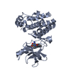
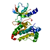
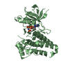
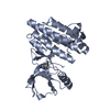
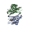
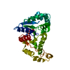
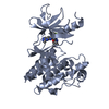
| Title | Crystal Structure of Ketosteroid Isomerase P39A from Pseudomonas Testosteroni (tKSI) | ||||||
 Components Components | Steroid Delta-Isomerase | ||||||
 Keywords Keywords | ISOMERASE / Steroid Metabolism | ||||||
| Function / homology |  Function and homology information Function and homology informationsteroid Delta-isomerase / steroid Delta-isomerase activity / steroid metabolic process Similarity search - Function | ||||||
| Biological species |  Comamonas testosteroni (bacteria) Comamonas testosteroni (bacteria) | ||||||
| Method |  X-RAY DIFFRACTION / SYNCHROTRON / MOLECULAR REPLACEMENT / molecular replacement / Resolution: 1.722 Å X-RAY DIFFRACTION / SYNCHROTRON / MOLECULAR REPLACEMENT / molecular replacement / Resolution: 1.722 Å | ||||||
 Authors Authors | Gonzalez, A. / Tsai, Y. / Schwans, J. / Sunden, F. / Herschlag, D. | ||||||
 Citation Citation | Journal: To be Published Title: Crystal Structure of Ketosteroid Isomerase P39A from Pseudomonas Testosteroni (tKSI) Authors: Schwans, J. / Sunden, F. / Gonzalez, A. / Tsai, Y. / Herschlag, D. | ||||||
| History |
|
- Structure visualization
| Structure viewer | Molecule: MolmilJmol/JSmol |
|---|
- Downloads & links
-Download
| PDBx/mmCIF format | 3mhe.cif.gz | 113.2 KB | Display | PDBx/mmCIF format |
|---|---|---|---|---|
| PDB format | pdb3mhe.ent.gz | 89.6 KB | Display | PDB format |
| PDBx/mmJSON format | 3mhe.json.gz | Tree view | PDBx/mmJSON format | |
| Others |  Other downloads Other downloads |
-Validation report
| Arichive directory | https://data.pdbj.org/pub/pdb/validation_reports/mh/3mheftp://data.pdbj.org/pub/pdb/validation_reports/mh/3mhe | HTTPS FTP |
|---|
-Related structure data
| Related structure data |  8choS S: Starting model for refinement |
|---|---|
| Similar structure data |


-Links
 PDBj
PDBj
- Assembly
| Deposited unit | 
| ||||||||
|---|---|---|---|---|---|---|---|---|---|
| 1 |
| ||||||||
| Unit cell |
|
-Components
| #1: Protein | Mass: 13386.064 Da / Num. of mol.: 2 / Mutation: P39A Source method: isolated from a genetically manipulated source Source: (gene. exp.) Comamonas testosteroni (bacteria) / Strain: ATCC 11996 / Gene: ksi / Plasmid: pKK / Production host: #2: Chemical | ChemComp-SO4 /   Mass: 96.063 Da / Num. of mol.: 7 / Source method: obtained synthetically / Formula: SO4 Mass: 96.063 Da / Num. of mol.: 7 / Source method: obtained synthetically / Formula: SO4#3: Chemical | ChemComp-POL / |   Mass: 60.095 Da / Num. of mol.: 1 / Source method: obtained synthetically / Formula: C3H8O Mass: 60.095 Da / Num. of mol.: 1 / Source method: obtained synthetically / Formula: C3H8O#4: Water | ChemComp-HOH / |  Mass: 18.015 Da / Num. of mol.: 156 / Source method: isolated from a natural source / Formula: H2O Mass: 18.015 Da / Num. of mol.: 156 / Source method: isolated from a natural source / Formula: H2O |
|---|
-Experimental details
-Experiment
| Experiment | Method: X-RAY DIFFRACTION / Number of used crystals: 1 |
|---|
- Sample preparation
| Crystal | Density Matthews: 2.18 Å3/Da / Density % sol: 43.7 % |
|---|---|
| Crystal grow | Temperature: 298 K / Method: vapor diffusion, sitting drop / pH: 7.2 Details: 2.0 M ammonium sulfate, 5% propanol, 20 mM potassium phosphate, 1 mM EDTA, 2 mM DTT, pH 7.2, vapor diffusion, sitting drop, temperature 298K, VAPOR DIFFUSION, SITTING DROP |
-Data collection
| Diffraction |
| ||||||||||||||||||
|---|---|---|---|---|---|---|---|---|---|---|---|---|---|---|---|---|---|---|---|
| Diffraction source |
| ||||||||||||||||||
| Detector |
| ||||||||||||||||||
| Radiation |
| ||||||||||||||||||
| Radiation wavelength | Wavelength: 0.98 Å / Relative weight: 1 | ||||||||||||||||||
| Reflection | Resolution: 1.722→29.732 Å / Num. all: 25533 / Num. obs: 25533 / % possible obs: 99.7 % / Observed criterion σ(F): 0 / Observed criterion σ(I): 0 / Redundancy: 13.6 % / Biso Wilson estimate: 23.8 Å2 / Rsym value: 0.046 / Net I/σ(I): 37.3 | ||||||||||||||||||
| Reflection shell | Resolution: 1.722→1.81 Å / Redundancy: 10.6 % / Mean I/σ(I) obs: 4.4 / Num. unique all: 38331 / Rsym value: 0.539 / % possible all: 100 |

-Phasing
| Phasing | Method: molecular replacement |
|---|
- Processing
| Software |
| |||||||||||||||||||||||||||||||||||||||||||||||||||||||||||||||||||||||||||
|---|---|---|---|---|---|---|---|---|---|---|---|---|---|---|---|---|---|---|---|---|---|---|---|---|---|---|---|---|---|---|---|---|---|---|---|---|---|---|---|---|---|---|---|---|---|---|---|---|---|---|---|---|---|---|---|---|---|---|---|---|---|---|---|---|---|---|---|---|---|---|---|---|---|---|---|---|
| Refinement | Method to determine structure: MOLECULAR REPLACEMENT Starting model: PDB ENTRY 8CHO Resolution: 1.722→29.73 Å / Cor.coef. Fo:Fc: 0.957 / Cor.coef. Fo:Fc free: 0.933 / WRfactor Rfree: 0.227 / WRfactor Rwork: 0.182 / Occupancy max: 1 / Occupancy min: 0.38 / FOM work R set: 0.873 / SU B: 5.12 / SU ML: 0.076 / SU R Cruickshank DPI: 0.123 / SU Rfree: 0.122 / Cross valid method: THROUGHOUT / σ(F): 0 / ESU R: 0.123 / ESU R Free: 0.122 / Stereochemistry target values: MAXIMUM LIKELIHOOD
| |||||||||||||||||||||||||||||||||||||||||||||||||||||||||||||||||||||||||||
| Solvent computation | Ion probe radii: 0.8 Å / Shrinkage radii: 0.8 Å / VDW probe radii: 1.4 Å / Solvent model: BABINET MODEL WITH MASK | |||||||||||||||||||||||||||||||||||||||||||||||||||||||||||||||||||||||||||
| Displacement parameters | Biso max: 116.42 Å2 / Biso mean: 26.212 Å2 / Biso min: 5.73 Å2
| |||||||||||||||||||||||||||||||||||||||||||||||||||||||||||||||||||||||||||
| Refinement step | Cycle: LAST / Resolution: 1.722→29.73 Å
| |||||||||||||||||||||||||||||||||||||||||||||||||||||||||||||||||||||||||||
| Refine LS restraints |
| |||||||||||||||||||||||||||||||||||||||||||||||||||||||||||||||||||||||||||
| LS refinement shell | Resolution: 1.722→1.766 Å / Total num. of bins used: 20
| |||||||||||||||||||||||||||||||||||||||||||||||||||||||||||||||||||||||||||
| Refinement TLS params. | Method: refined / Refine-ID: X-RAY DIFFRACTION
| |||||||||||||||||||||||||||||||||||||||||||||||||||||||||||||||||||||||||||
| Refinement TLS group |
|
