 Movie
Movie Controller

[English] 日本語
 Yorodumi
- PDB-3gay: Structure of Giardia fructose-1,6-biphosphate aldolase in complex... -
+ Open data

- Basic information
| Entry | Database: PDB / ID: 3gay | ||||||
|---|---|---|---|---|---|---|---|
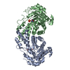
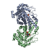
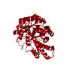
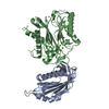
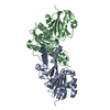
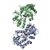
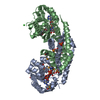
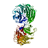
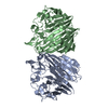
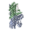
| Title | Structure of Giardia fructose-1,6-biphosphate aldolase in complex with tagatose-1,6-biphosphate | ||||||
 Components Components | Fructose-bisphosphate aldolase | ||||||
 Keywords Keywords | LYASE / Class II fructose-1 / 6-bisphosphate aldolase / glycolytic pathway / Giardia lamblia / drug target / Glycolysis | ||||||
| Function / homology |  Function and homology information Function and homology informationfructose-bisphosphate aldolase / fructose-bisphosphate aldolase activity / fructose 1,6-bisphosphate metabolic process / glycolytic process / zinc ion binding Similarity search - Function | ||||||
| Biological species |  Giardia intestinalis (eukaryote) Giardia intestinalis (eukaryote) | ||||||
| Method |  X-RAY DIFFRACTION / MOLECULAR REPLACEMENT / Resolution: 1.8 Å X-RAY DIFFRACTION / MOLECULAR REPLACEMENT / Resolution: 1.8 Å | ||||||
 Authors Authors | Galkin, A. / Herzberg, O. | ||||||
 Citation Citation | Journal: Biochemistry / Year: 2009 Title: Structural insights into the substrate binding and stereoselectivity of giardia fructose-1,6-bisphosphate aldolase. Authors: Galkin, A. / Li, Z. / Li, L. / Kulakova, L. / Pal, L.R. / Dunaway-Mariano, D. / Herzberg, O. | ||||||
| History |
|
- Structure visualization
| Structure viewer | Molecule: MolmilJmol/JSmol |
|---|
- Downloads & links
-Download
| PDBx/mmCIF format | 3gay.cif.gz | 146.3 KB | Display | PDBx/mmCIF format |
|---|---|---|---|---|
| PDB format | pdb3gay.ent.gz | 113.1 KB | Display | PDB format |
| PDBx/mmJSON format | 3gay.json.gz | Tree view | PDBx/mmJSON format | |
| Others |  Other downloads Other downloads |
-Validation report
| Arichive directory | https://data.pdbj.org/pub/pdb/validation_reports/ga/3gayftp://data.pdbj.org/pub/pdb/validation_reports/ga/3gay | HTTPS FTP |
|---|
-Related structure data
| Related structure data |  3gakC  3gb6C 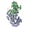 2iswS S: Starting model for refinement C: citing same article ( |
|---|---|
| Similar structure data |

-Links
 PDBj
PDBj
- Assembly
| Deposited unit | 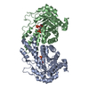
| ||||||||
|---|---|---|---|---|---|---|---|---|---|
| 1 |
| ||||||||
| Unit cell |
|
-Components
| #1: Protein | Mass: 35263.758 Da / Num. of mol.: 2 Source method: isolated from a genetically manipulated source Source: (gene. exp.) Giardia intestinalis (eukaryote) / Strain: WB / Gene: ald, FBPA / Plasmid: pET100/D-TOPO / Production host:  #2: Chemical |   Mass: 65.409 Da / Num. of mol.: 2 / Source method: obtained synthetically / Formula: Zn Mass: 65.409 Da / Num. of mol.: 2 / Source method: obtained synthetically / Formula: Zn#3: Chemical |   Mass: 340.116 Da / Num. of mol.: 2 / Source method: obtained synthetically / Formula: C6H14O12P2 Mass: 340.116 Da / Num. of mol.: 2 / Source method: obtained synthetically / Formula: C6H14O12P2#4: Water | ChemComp-HOH / |  Mass: 18.015 Da / Num. of mol.: 578 / Source method: isolated from a natural source / Formula: H2O Mass: 18.015 Da / Num. of mol.: 578 / Source method: isolated from a natural source / Formula: H2OSequence details | AUTHORS STATE THAT ACCORDING TO THE SEQUENCE FROM GIARDIA GENOME (GB ENTRY EAA46366.1) THE CORRECT ...AUTHORS STATE THAT ACCORDING TO THE SEQUENCE FROM GIARDIA GENOME (GB ENTRY EAA46366.1) THE CORRECT RESIDUE AT POSITION 129 IS GLY. | |
|---|
-Experimental details
-Experiment
| Experiment | Method: X-RAY DIFFRACTION / Number of used crystals: 1 |
|---|
- Sample preparation
| Crystal | Density Matthews: 2.31 Å3/Da / Density % sol: 46.86 % |
|---|---|
| Crystal grow | Temperature: 298 K / Method: vapor diffusion, hanging drop / pH: 7.5 Details: 18-25% PEG3350, 0.2M NH4NO3, pH 7.5, VAPOR DIFFUSION, HANGING DROP, temperature 298.0K |
-Data collection
| Diffraction | Mean temperature: 100 K |
|---|---|
| Diffraction source | Source: ROTATING ANODE / Type: RIGAKU / Wavelength: 1.54178 Å |
| Detector | Type: RIGAKU RAXIS IV++ / Detector: IMAGE PLATE / Date: Apr 9, 2007 / Details: mirrors |
| Radiation | Monochromator: OSMIC MIRRORS / Protocol: SINGLE WAVELENGTH / Monochromatic (M) / Laue (L): M / Scattering type: x-ray |
| Radiation wavelength | Wavelength: 1.54178 Å / Relative weight: 1 |
| Reflection | Resolution: 1.8→20 Å / Num. all: 61609 / Num. obs: 57851 / % possible obs: 93.9 % / Rmerge(I) obs: 0.066 |
| Reflection shell | Resolution: 1.8→1.86 Å / Rmerge(I) obs: 0.342 / % possible all: 62.9 |
- Processing
| Software |
| |||||||||||||||||||||||||
|---|---|---|---|---|---|---|---|---|---|---|---|---|---|---|---|---|---|---|---|---|---|---|---|---|---|---|
| Refinement | Method to determine structure: MOLECULAR REPLACEMENT Starting model: PDB ENTRY 2ISW Resolution: 1.8→20 Å / Isotropic thermal model: Isotropic / Cross valid method: THROUGHOUT / σ(F): 0 / Stereochemistry target values: Engh & Huber
| |||||||||||||||||||||||||
| Refinement step | Cycle: LAST / Resolution: 1.8→20 Å
| |||||||||||||||||||||||||
| Refine LS restraints |
|
