 Movie
Movie Controller

[English] 日本語
 Yorodumi
- PDB-2isv: Structure of Giardia fructose-1,6-biphosphate aldolase in complex... -
+ Open data

- Basic information
| Entry | Database: PDB / ID: 2isv | ||||||
|---|---|---|---|---|---|---|---|
| Title | Structure of Giardia fructose-1,6-biphosphate aldolase in complex with phosphoglycolohydroxamate | ||||||
 Components Components | Putative fructose-1,6-bisphosphate aldolase | ||||||
 Keywords Keywords | LYASE / Class II fructose-1 / 6-bisphosphate aldolase / glycolytic pathway / Giardia lamblia / drug target | ||||||
| Function / homology |  Function and homology information Function and homology informationfructose-bisphosphate aldolase / fructose-bisphosphate aldolase activity / fructose 1,6-bisphosphate metabolic process / glycolytic process / zinc ion binding Similarity search - Function | ||||||
| Biological species |  Giardia intestinalis (eukaryote) Giardia intestinalis (eukaryote) | ||||||
| Method |  X-RAY DIFFRACTION / SYNCHROTRON / MOLECULAR REPLACEMENT / Resolution: 2.3 Å X-RAY DIFFRACTION / SYNCHROTRON / MOLECULAR REPLACEMENT / Resolution: 2.3 Å | ||||||
 Authors Authors | Galkin, A. / Herzberg, O. | ||||||
 Citation Citation | Journal: J.Biol.Chem. / Year: 2007 Title: Characterization, kinetics, and crystal structures of fructose-1,6-bisphosphate aldolase from the human parasite, Giardia lamblia. Authors: Galkin, A. / Kulakova, L. / Melamud, E. / Li, L. / Wu, C. / Mariano, P. / Dunaway-Mariano, D. / Nash, T.E. / Herzberg, O. | ||||||
| History |
|
- Structure visualization
| Structure viewer | Molecule: MolmilJmol/JSmol |
|---|
- Downloads & links
-Download
| PDBx/mmCIF format | 2isv.cif.gz | 135.8 KB | Display | PDBx/mmCIF format |
|---|---|---|---|---|
| PDB format | pdb2isv.ent.gz | 105.6 KB | Display | PDB format |
| PDBx/mmJSON format | 2isv.json.gz | Tree view | PDBx/mmJSON format | |
| Others |  Other downloads Other downloads |
-Validation report
| Arichive directory | https://data.pdbj.org/pub/pdb/validation_reports/is/2isvftp://data.pdbj.org/pub/pdb/validation_reports/is/2isv | HTTPS FTP |
|---|
-Related structure data
| Related structure data | 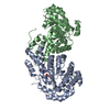 2iswC  1jvf C: citing same article ( S: Starting model for refinement |
|---|---|
| Similar structure data |


-Links
 PDBj
PDBj
- Assembly
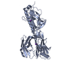
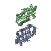
| Deposited unit | 
| ||||||||
|---|---|---|---|---|---|---|---|---|---|
| 1 |
| ||||||||
| Unit cell |
| ||||||||
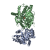
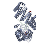
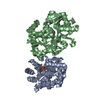
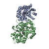
| Details | The biological assembly is a dimer generated from the molecules A and B in the asymmetric unit |
-Components
| #1: Protein | Mass: 35293.785 Da / Num. of mol.: 2 Source method: isolated from a genetically manipulated source Source: (gene. exp.) Giardia intestinalis (eukaryote) / Strain: WB / Gene: ald / Plasmid: pET100/FBPAn / Production host:  #2: Chemical |   Mass: 65.409 Da / Num. of mol.: 2 / Source method: obtained synthetically / Formula: Zn Mass: 65.409 Da / Num. of mol.: 2 / Source method: obtained synthetically / Formula: Zn#3: Chemical | 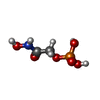  Mass: 171.046 Da / Num. of mol.: 2 / Source method: obtained synthetically / Formula: C2H6NO6P Mass: 171.046 Da / Num. of mol.: 2 / Source method: obtained synthetically / Formula: C2H6NO6P#4: Water | ChemComp-HOH / |  Mass: 18.015 Da / Num. of mol.: 368 / Source method: isolated from a natural source / Formula: H2O Mass: 18.015 Da / Num. of mol.: 368 / Source method: isolated from a natural source / Formula: H2O |
|---|
-Experimental details
-Experiment
| Experiment | Method: X-RAY DIFFRACTION / Number of used crystals: 1 |
|---|
- Sample preparation
| Crystal | Density Matthews: 2.54 Å3/Da / Density % sol: 51.62 % |
|---|---|
| Crystal grow | Temperature: 298 K / Method: vapor diffusion, hanging drop / pH: 8.75 Details: 18-23% Polyethylglycol monomethyl ether 2000, 0.1 M Tris-HCl, 0.2 M MgCl2, 20 mM PGH, pH 8.75, VAPOR DIFFUSION, HANGING DROP, temperature 298.0K |
-Data collection
| Diffraction | Mean temperature: 100 K |
|---|---|
| Diffraction source | Source: SYNCHROTRON / Site: APS  / Beamline: 17-ID / Wavelength: 1 Å / Beamline: 17-ID / Wavelength: 1 Å |
| Detector | Type: ADSC QUANTUM 210 / Detector: CCD / Date: Jul 7, 2003 |
| Radiation | Monochromator: Si 111 CHANNEL / Protocol: SINGLE WAVELENGTH / Monochromatic (M) / Laue (L): M / Scattering type: x-ray |
| Radiation wavelength | Wavelength: 1 Å / Relative weight: 1 |
| Reflection | Resolution: 2.3→30 Å / Num. all: 31046 / Num. obs: 28450 / % possible obs: 91.6 % / Rmerge(I) obs: 0.037 |
| Reflection shell | Resolution: 2.3→2.38 Å / Rmerge(I) obs: 0.116 / % possible all: 77.9 |
- Processing
| Software |
| |||||||||||||||||||||||||
|---|---|---|---|---|---|---|---|---|---|---|---|---|---|---|---|---|---|---|---|---|---|---|---|---|---|---|
| Refinement | Method to determine structure: MOLECULAR REPLACEMENT Starting model: PDB ENTRY 1JVF 1jvf Resolution: 2.3→30 Å / Isotropic thermal model: Isotropic / Cross valid method: THROUGHOUT / σ(F): 0 / Stereochemistry target values: Engh & Huber
| |||||||||||||||||||||||||
| Displacement parameters | Biso mean: 51.3 Å2 | |||||||||||||||||||||||||
| Refinement step | Cycle: LAST / Resolution: 2.3→30 Å
| |||||||||||||||||||||||||
| Refine LS restraints |
| |||||||||||||||||||||||||
| LS refinement shell | Resolution: 2.3→2.38 Å
|
