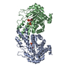
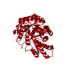
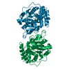
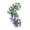
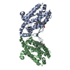
登録情報 データベース : PDB / ID : 1nr0タイトル Two Seven-Bladed Beta-Propeller Domains Revealed By The Structure Of A C. elegans Homologue Of Yeast Actin Interacting Protein 1 (AIP1). Actin interacting protein 1 キーワード / / / / / / / / / / 機能・相同性 分子機能 ドメイン・相同性 構成要素
/ / / / / / / / / / / / / / / / / / / / / / / / / / / / / / / / / / / / / / / / / / / 生物種 Caenorhabditis elegans (センチュウ)手法 / / / 解像度 : 1.7 Å データ登録者 Vorobiev, S.M. / Mohri, K. / Ono, S. / Almo, S.C. / Burley, S.K. / New York SGX Research Center for Structural Genomics (NYSGXRC) ジャーナル : J.Biol.Chem. / 年 : 2004タイトル : Identification of Functional Residues on Caenorhabditis elegans Actin-interacting Protein 1 (UNC-78) for Disassembly of Actin Depolymerizing Factor/Cofilin-bound Actin Filaments著者 : Mohri, K. / Vorobiev, S.M. / Fedorov, A.A. / Almo, S.C. / Ono, S. 履歴 登録 2003年1月23日 登録サイト / 処理サイト 改定 1.0 2003年7月1日 Provider / タイプ 改定 1.1 2008年4月29日 Group 改定 1.2 2011年7月13日 Group 改定 1.3 2021年2月3日 Group / Structure summaryカテゴリ audit_author / pdbx_struct_conn_angle ... audit_author / pdbx_struct_conn_angle / struct_conn / struct_site Item _audit_author.identifier_ORCID / _pdbx_struct_conn_angle.ptnr1_auth_seq_id ... _audit_author.identifier_ORCID / _pdbx_struct_conn_angle.ptnr1_auth_seq_id / _pdbx_struct_conn_angle.ptnr3_auth_seq_id / _pdbx_struct_conn_angle.value / _struct_conn.pdbx_dist_value / _struct_conn.ptnr1_auth_comp_id / _struct_conn.ptnr1_auth_seq_id / _struct_conn.ptnr1_label_asym_id / _struct_conn.ptnr1_label_atom_id / _struct_conn.ptnr1_label_comp_id / _struct_conn.ptnr1_label_seq_id / _struct_conn.ptnr2_auth_comp_id / _struct_conn.ptnr2_auth_seq_id / _struct_conn.ptnr2_label_asym_id / _struct_conn.ptnr2_label_atom_id / _struct_conn.ptnr2_label_comp_id / _struct_conn.ptnr2_label_seq_id / _struct_site.pdbx_auth_asym_id / _struct_site.pdbx_auth_comp_id / _struct_site.pdbx_auth_seq_id 改定 1.4 2024年2月14日 Group / Database references / カテゴリ / chem_comp_bond / database_2Item / _database_2.pdbx_database_accession
すべて表示 表示を減らす
 ムービー
ムービー コントローラー


 データを開く
 基本情報
基本情報 要素
要素 キーワード
キーワード 機能・相同性情報
 X線回折 /
  データ登録者
 引用
引用 構造の表示
構造の表示 ダウンロードとリンク
 その他のダウンロード


 PDBj
PDBj 集合体
集合体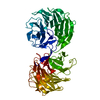


 分子量: 54.938 Da / 分子数: 1 / 由来タイプ: 合成 / 式: Mn
 分子量: 18.015 Da / 分子数: 709 / 由来タイプ: 天然 / 式: H2O
 試料調製
試料調製 / ビームライン: X9B / 波長: 0.98 Å
 解析
解析