Mass: 18.015 Da / Num. of mol.: 359 / Source method: isolated from a natural source / Formula: H2O
Has protein modification
Y
Sequence details
THIS CONSTRUCT WAS EXPRESSED WITH A PURIFICATION TAG MGSDKIHHHHHHENLYFQG. THE TAG WAS REMOVED WITH ...THIS CONSTRUCT WAS EXPRESSED WITH A PURIFICATION TAG MGSDKIHHHHHHENLYFQG. THE TAG WAS REMOVED WITH TEV PROTEASE LEAVING ONLY A GLYCINE (0) FOLLOWED BY THE TARGET SEQUENCE.
-
Experimental details
-
Experiment
Experiment
Method: X-RAY DIFFRACTION / Number of used crystals: 1
-
Sample preparation
Crystal
Density Matthews: 2.49 Å3/Da / Density % sol: 50.54 %
Crystal grow
Temperature: 277 K / Method: vapor diffusion, sitting drop / pH: 9.1 Details: NANODROP, 0.20M Na2HPO4, 20.0% PEG 3350, No Buffer pH 9.1, VAPOR DIFFUSION, SITTING DROP, temperature 277K
Type: MARMOSAIC 325 mm CCD / Detector: CCD / Date: Nov 13, 2008 / Details: Flat mirror (vertical focusing)
Radiation
Monochromator: Single crystal Si(111) bent (horizontal focusing) Protocol: MAD / Monochromatic (M) / Laue (L): M / Scattering type: x-ray
Radiation wavelength
ID
Wavelength (Å)
Relative weight
1
0.91162
1
2
0.97882
1
3
0.97828
1
Reflection
Resolution: 2.1→29.828 Å / Num. obs: 36707 / % possible obs: 99.9 % / Redundancy: 3.7 % / Biso Wilson estimate: 27.96 Å2 / Rmerge(I) obs: 0.108 / Rsym value: 0.108 / Net I/σ(I): 10.4
Reflection shell
Diffraction-ID: 1
Resolution (Å)
Redundancy (%)
Rmerge(I) obs
Mean I/σ(I) obs
Num. measured all
Num. unique all
Rsym value
% possible all
2.1-2.15
3.6
0.503
2.1
9838
2704
0.503
99.6
2.15-2.21
3.7
0.449
2.4
9609
2614
0.449
99.8
2.21-2.28
3.7
0.364
2.9
9318
2525
0.364
99.9
2.28-2.35
3.7
0.345
3.2
9227
2510
0.345
99.9
2.35-2.42
3.7
0.282
4
8965
2416
0.282
100
2.42-2.51
3.7
0.253
4.6
8619
2308
0.253
99.9
2.51-2.6
3.7
0.233
4.9
8471
2260
0.233
100
2.6-2.71
3.8
0.181
6.4
8215
2163
0.181
100
2.71-2.83
3.8
0.16
7.6
7996
2086
0.16
100
2.83-2.97
3.8
0.134
9.3
7676
1995
0.134
100
2.97-3.13
3.8
0.11
11.9
7241
1885
0.11
100
3.13-3.32
3.8
0.095
14.9
6921
1808
0.095
100
3.32-3.55
3.8
0.081
18
6500
1701
0.081
100
3.55-3.83
3.8
0.068
22
5978
1566
0.068
100
3.83-4.2
3.8
0.063
25
5562
1457
0.063
100
4.2-4.7
3.8
0.058
26.4
4967
1315
0.058
100
4.7-5.42
3.8
0.059
26.4
4443
1182
0.059
100
5.42-6.64
3.7
0.067
24.2
3677
992
0.067
100
6.64-9.39
3.6
0.07
27.5
2798
785
0.07
100
9.39-29.828
3.2
0.07
32.2
1378
435
0.07
96.9
-
Phasing
Phasing
Method: MAD
-
Processing
Software
Name
Version
Classification
NB
REFMAC
5.2.0019
refinement
PHENIX
refinement
SHELX
phasing
MolProbity
3beta29
modelbuilding
SCALA
3.2.5
datascaling
PDB_EXTRACT
3.006
dataextraction
MAR345
CCD
datacollection
MOSFLM
datareduction
SHELXD
phasing
autoSHARP
phasing
Refinement
Method to determine structure: MAD / Resolution: 2.1→29.828 Å / Cor.coef. Fo:Fc: 0.954 / Cor.coef. Fo:Fc free: 0.928 / Occupancy max: 1 / Occupancy min: 0.2 / SU B: 9.981 / SU ML: 0.138 / TLS residual ADP flag: LIKELY RESIDUAL / Cross valid method: THROUGHOUT / σ(F): 0 / ESU R: 0.216 / ESU R Free: 0.18 Stereochemistry target values: MAXIMUM LIKELIHOOD WITH PHASES Details: 1. HYDROGENS HAVE BEEN ADDED IN THE RIDING POSITIONS. 2. ATOM RECORDS CONTAIN RESIDUAL B FACTORS ONLY. 3. A MET-INHIBITION PROTOCOL WAS USED FOR SELENOMETHIONINE INCORPORATION DURING PROTEIN ...Details: 1. HYDROGENS HAVE BEEN ADDED IN THE RIDING POSITIONS. 2. ATOM RECORDS CONTAIN RESIDUAL B FACTORS ONLY. 3. A MET-INHIBITION PROTOCOL WAS USED FOR SELENOMETHIONINE INCORPORATION DURING PROTEIN EXPRESSION. THE OCCUPANCY OF THE SE ATOMS IN THE MSE RESIDUES WAS REDUCED TO 0.75 FOR THE REDUCED SCATTERING POWER DUE TO PARTIAL S-MET INCORPORATION. 4. TLS GROUPS WERE ASSIGNED WITH THE AID OF TLSMD. 5. NA ION, ETHYLENE GLYCOL AND PHOSPHATE MOLECULES FROM THE CRYSTALLIZATION/CRYO SOLUTION ARE MODELED.
Rfactor
Num. reflection
% reflection
Selection details
Rfree
0.23
1833
5 %
RANDOM
Rwork
0.188
-
-
-
obs
0.19
36695
99.79 %
-
Solvent computation
Ion probe radii: 0.8 Å / Shrinkage radii: 0.8 Å / VDW probe radii: 1.2 Å / Solvent model: BABINET MODEL WITH MASK
In the structure databanks used in Yorodumi, some data are registered as the other names, "COVID-19 virus" and "2019-nCoV". Here are the details of the virus and the list of structure data.
Jan 31, 2019. EMDB accession codes are about to change! (news from PDBe EMDB page)
EMDB accession codes are about to change! (news from PDBe EMDB page)
The allocation of 4 digits for EMDB accession codes will soon come to an end. Whilst these codes will remain in use, new EMDB accession codes will include an additional digit and will expand incrementally as the available range of codes is exhausted. The current 4-digit format prefixed with “EMD-” (i.e. EMD-XXXX) will advance to a 5-digit format (i.e. EMD-XXXXX), and so on. It is currently estimated that the 4-digit codes will be depleted around Spring 2019, at which point the 5-digit format will come into force.
The EM Navigator/Yorodumi systems omit the EMD- prefix.
Related info.:Q: What is EMD? / ID/Accession-code notation in Yorodumi/EM Navigator
Yorodumi is a browser for structure data from EMDB, PDB, SASBDB, etc.
This page is also the successor to EM Navigator detail page, and also detail information page/front-end page for Omokage search.
The word "yorodu" (or yorozu) is an old Japanese word meaning "ten thousand". "mi" (miru) is to see.
Related info.:EMDB / PDB / SASBDB / Comparison of 3 databanks / Yorodumi Search / Aug 31, 2016. New EM Navigator & Yorodumi / Yorodumi Papers / Jmol/JSmol / Function and homology information / Changes in new EM Navigator and Yorodumi
 Movie
Movie Controller

 Yorodumi
 Open data
 Basic information
 Components
 Keywords
 Function and homology information
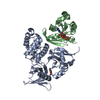
 Clostridium difficile (bacteria)
 X-RAY DIFFRACTION /
  Authors
 Citation
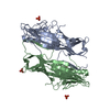
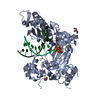
 Structure visualization
 Downloads & links
 Other downloads
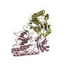


 PDBj
PDBj

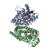
 Assembly
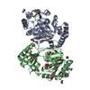

 Mass: 94.971 Da / Num. of mol.: 2 / Source method: obtained synthetically / Formula: PO4
 Mass: 62.068 Da / Num. of mol.: 8 / Source method: obtained synthetically / Formula: C2H6O2
 Mass: 22.990 Da / Num. of mol.: 1 / Source method: obtained synthetically / Formula: Na
 Mass: 18.015 Da / Num. of mol.: 359 / Source method: isolated from a natural source / Formula: H2O
 Sample preparation
 / Beamline: BL11-1 / Wavelength: 0.91162, 0.97882, 0.97828
 Processing