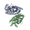
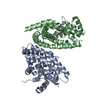
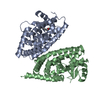
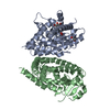
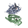
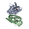
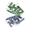
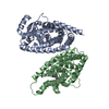
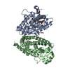
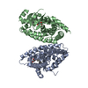
[English] 日本語
Yorodumi
- PDB-3wmh: Human PPAR gamma ligand binding domain in complex with a gammma s... -

+
Open data


ID or keywords:

Loading...

-
Basic information

Entry
Database: PDB / ID: 3wmh
TitleHuman PPAR gamma ligand binding domain in complex with a gammma selective synthetic partial agonist MEKT75
ComponentsPeroxisome proliferator-activated receptor gamma
KeywordsTRANSCRIPTION / Mainly Alpha / Nuclear Receptor
Function / homology
Function and homology information


prostaglandin receptor activity / : / negative regulation of receptor signaling pathway via STAT / MECP2 regulates transcription factors / negative regulation of vascular endothelial cell proliferation / negative regulation of extracellular matrix assembly / negative regulation of connective tissue replacement involved in inflammatory response wound healing / positive regulation of cholesterol transport / negative regulation of cellular response to transforming growth factor beta stimulus / arachidonate binding ...prostaglandin receptor activity / : / negative regulation of receptor signaling pathway via STAT / MECP2 regulates transcription factors / negative regulation of vascular endothelial cell proliferation / negative regulation of extracellular matrix assembly / negative regulation of connective tissue replacement involved in inflammatory response wound healing / positive regulation of cholesterol transport / negative regulation of cellular response to transforming growth factor beta stimulus / arachidonate binding / positive regulation of adiponectin secretion / DNA binding domain binding / lipoprotein transport / negative regulation of cardiac muscle hypertrophy in response to stress / positive regulation of vascular associated smooth muscle cell apoptotic process / WW domain binding / positive regulation of fatty acid metabolic process / STAT family protein binding / response to lipid / negative regulation of SMAD protein signal transduction / negative regulation of type II interferon-mediated signaling pathway / LBD domain binding / negative regulation of cholesterol storage / lipid homeostasis / E-box binding / alpha-actinin binding / R-SMAD binding / negative regulation of vascular associated smooth muscle cell proliferation / monocyte differentiation / negative regulation of blood vessel endothelial cell migration / white fat cell differentiation / negative regulation of macrophage derived foam cell differentiation / negative regulation of lipid storage / cellular response to low-density lipoprotein particle stimulus / positive regulation of cholesterol efflux / negative regulation of BMP signaling pathway / cell fate commitment / negative regulation of mitochondrial fission / negative regulation of osteoblast differentiation / positive regulation of fat cell differentiation / BMP signaling pathway / long-chain fatty acid transport / nuclear retinoid X receptor binding / Transcriptional regulation of brown and beige adipocyte differentiation by EBF2 / retinoic acid receptor signaling pathway / cell maturation / negative regulation of MAPK cascade / intracellular receptor signaling pathway / hormone-mediated signaling pathway / positive regulation of adipose tissue development / peroxisome proliferator activated receptor signaling pathway / epithelial cell differentiation / regulation of cellular response to insulin stimulus / response to nutrient / peptide binding / negative regulation of miRNA transcription / placenta development / negative regulation of angiogenesis / transcription coregulator binding / Regulation of PTEN gene transcription / positive regulation of apoptotic signaling pathway / SUMOylation of intracellular receptors / negative regulation of smooth muscle cell proliferation / negative regulation of transforming growth factor beta receptor signaling pathway / fatty acid metabolic process / mRNA transcription by RNA polymerase II / PPARA activates gene expression / regulation of circadian rhythm / Transcriptional regulation of white adipocyte differentiation / Nuclear Receptor transcription pathway / regulation of blood pressure / lipid metabolic process / positive regulation of miRNA transcription / negative regulation of inflammatory response / DNA-binding transcription repressor activity, RNA polymerase II-specific / RNA polymerase II transcription regulator complex / cellular response to insulin stimulus / nuclear receptor activity / rhythmic process / glucose homeostasis / MLL4 and MLL3 complexes regulate expression of PPARG target genes in adipogenesis and hepatic steatosis / DNA-binding transcription activator activity, RNA polymerase II-specific / double-stranded DNA binding / cellular response to hypoxia / DNA-binding transcription factor binding / sequence-specific DNA binding / nucleic acid binding / cell differentiation / DNA-binding transcription factor activity, RNA polymerase II-specific / receptor complex / transcription cis-regulatory region binding / RNA polymerase II cis-regulatory region sequence-specific DNA binding / DNA-binding transcription factor activity / negative regulation of gene expression / innate immune response / negative regulation of DNA-templated transcription / intracellular membrane-bounded organelle / chromatin binding / positive regulation of gene expression / regulation of transcription by RNA polymerase II
Similarity search - Function
Peroxisome proliferator-activated receptor gamma / Peroxisome proliferator-activated receptor gamma, N-terminal / PPAR gamma N-terminal region / Peroxisome proliferator-activated receptor / : / Retinoid X Receptor / Retinoid X Receptor / Nuclear hormone receptor / Nuclear hormones receptors DNA-binding region signature. / Zinc finger, nuclear hormone receptor-type ...Peroxisome proliferator-activated receptor gamma / Peroxisome proliferator-activated receptor gamma, N-terminal / PPAR gamma N-terminal region / Peroxisome proliferator-activated receptor / : / Retinoid X Receptor / Retinoid X Receptor / Nuclear hormone receptor / Nuclear hormones receptors DNA-binding region signature. / Zinc finger, nuclear hormone receptor-type / Double treble clef zinc finger, C4 type / Nuclear hormone receptors DNA-binding domain profile. / c4 zinc finger in nuclear hormone receptors / Nuclear hormone receptor, ligand-binding domain / Nuclear hormone receptor-like domain superfamily / Ligand-binding domain of nuclear hormone receptor / Nuclear receptor (NR) ligand-binding (LBD) domain profile. / Ligand binding domain of hormone receptors / Zinc finger, NHR/GATA-type / Orthogonal Bundle / Mainly Alpha
Similarity search - Domain/homology
Chem-JJA / Peroxisome proliferator-activated receptor gamma
Similarity search - Component
Biological speciesHomo sapiens (human)
MethodX-RAY DIFFRACTION / SYNCHROTRON / MOLECULAR REPLACEMENT / Resolution: 2.1 Å
AuthorsOyama, T. / Ohashi, M. / Miyachi, H. / Kusunoki, M.
CitationJournal: TO BE PUBLISHED
Title: Human PPRR gamma ligand binding domain in complex with a gammma selective synthetic partial agonist MEKT75
Authors: Oyama, T. / Ohashi, M. / Miyachi, H. / Kusunoki, M.
History
DepositionNov 19, 2013Deposition site: PDBJ / Processing site: PDBJ
Revision 1.0Nov 19, 2014Provider: repository / Type: Initial release
Revision 1.1Mar 2, 2016Group: Structure summary
Revision 1.2Nov 22, 2017Group: Refinement description / Category: software / Item: _software.name
Revision 1.3Nov 8, 2023Group: Data collection / Database references ...Data collection / Database references / Derived calculations / Refinement description
Category: chem_comp_atom / chem_comp_bond ...chem_comp_atom / chem_comp_bond / database_2 / pdbx_initial_refinement_model / struct_ref_seq_dif / struct_site
Item: _database_2.pdbx_DOI / _database_2.pdbx_database_accession ..._database_2.pdbx_DOI / _database_2.pdbx_database_accession / _struct_ref_seq_dif.details / _struct_site.pdbx_auth_asym_id / _struct_site.pdbx_auth_comp_id / _struct_site.pdbx_auth_seq_id

-
Structure visualization

Structure viewerMolecule:
MolmilJmol/JSmol

Downloads & links

-
Assembly

Deposited unit
A: Peroxisome proliferator-activated receptor gamma
B: Peroxisome proliferator-activated receptor gamma
hetero molecules


Theoretical massNumber of molelcules
Total (without water)66,1224
Polymers65,0612
Non-polymers1,0612
Water1,29772
1


  • Idetical with deposited unit
  • defined by author
TypeNameSymmetry operationNumber
identity operation1_555x,y,z1
Unit cell
Length a, b, c (Å)93.249, 61.113, 119.280
Angle α, β, γ (deg.)90.00, 103.11, 90.00
Int Tables number5
Space group name H-MC121

-
Components

#1: Protein Peroxisome proliferator-activated receptor gamma / PPAR-gamma / Nuclear receptor subfamily 1 group C member 3


Mass: 32530.652 Da / Num. of mol.: 2 / Fragment: Ligand binding domain, UNP residues 223-504
Source method: isolated from a genetically manipulated source
Source: (gene. exp.) Homo sapiens (human) / Gene: PPARG / Plasmid: pET28a / Production host: Escherichia coli (E. coli) / Strain (production host): BL21(DE3) / References: UniProt: P37231
#2: Chemical ChemComp-JJA / N-(phenylsulfonyl)-4-propoxy-3-({[4-(pyrimidin-2-yl)benzoyl]amino}methyl)benzamide


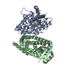
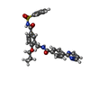
Mass: 530.595 Da / Num. of mol.: 2 / Source method: obtained synthetically / Formula: C28H26N4O5S
#3: Water ChemComp-HOH / water


Mass: 18.015 Da / Num. of mol.: 72 / Source method: isolated from a natural source / Formula: H2O

-
Experimental details

-
Experiment

ExperimentMethod: X-RAY DIFFRACTION / Number of used crystals: 1

-
Sample preparation

CrystalDensity Matthews: 2.54 Å3/Da / Density % sol: 51.65 %
Crystal growTemperature: 293 K / Method: vapor diffusion, hanging drop / pH: 7.5
Details: 0.8M SODIUM CITRATE, 0.1M HEPES, pH 7.5, VAPOR DIFFUSION, HANGING DROP, temperature 293K

-
Data collection

DiffractionMean temperature: 100 K
Diffraction sourceSource: SYNCHROTRON / Site: SPring-8 / Beamline: BL38B1 / Wavelength: 1 Å
DetectorType: ADSC QUANTUM 315 / Detector: CCD / Date: Jul 9, 2013
RadiationMonochromator: FIXED EXIT SI 111 DOUBLE CRYSTAL / Protocol: SINGLE WAVELENGTH / Monochromatic (M) / Laue (L): M / Scattering type: x-ray
Radiation wavelengthWavelength: 1 Å / Relative weight: 1
ReflectionResolution: 2.1→44.45 Å / Num. obs: 38348 / % possible obs: 98.8 % / Observed criterion σ(I): 0 / Redundancy: 5 % / Biso Wilson estimate: 42.4 Å2 / Rmerge(I) obs: 0.053 / Net I/σ(I): 19.7
Reflection shellResolution: 2.1→2.14 Å / Redundancy: 3.1 % / Rmerge(I) obs: 0.362 / Mean I/σ(I) obs: 2.68 / Num. unique all: 3780 / % possible all: 93.2

-
Processing

Software
NameVersionClassification
HKL-2000data collection
PHENIXmodel building
PHENIX(phenix.refine: 1.7.3_928)refinement
HKL-2000data reduction
HKL-2000data scaling
PHENIXphasing
RefinementMethod to determine structure: MOLECULAR REPLACEMENT
Starting model: PDB ENTRY 3VSO
Resolution: 2.1→44.45 Å / Occupancy max: 1 / Occupancy min: 1 / FOM work R set: 0.7734 / SU ML: 0.28 / Cross valid method: THROUGHOUT / σ(F): 1.34 / Phase error: 29.27 / Stereochemistry target values: ML
RfactorNum. reflection% reflectionSelection details
Rfree0.2575 1902 5.03 %random
Rwork0.2147 ---
obs0.2168 37839 98.56 %-
Solvent computationShrinkage radii: 0.98 Å / VDW probe radii: 1.2 Å / Solvent model: FLAT BULK SOLVENT MODEL / Bsol: 40.684 Å2 / ksol: 0.331 e/Å3
Displacement parametersBiso max: 109.98 Å2 / Biso mean: 54.4264 Å2 / Biso min: 29.46 Å2
Baniso -1Baniso -2Baniso -3
1--3.4936 Å20 Å24.3556 Å2
2--15.9144 Å20 Å2
3----12.4208 Å2
Refinement stepCycle: LAST / Resolution: 2.1→44.45 Å
ProteinNucleic acidLigandSolventTotal
Num. atoms4165 0 76 72 4313
Refine LS restraints
Refine-IDTypeDev idealNumber
X-RAY DIFFRACTIONf_bond_d0.0084317
X-RAY DIFFRACTIONf_angle_d0.9935814
X-RAY DIFFRACTIONf_chiral_restr0.065661
X-RAY DIFFRACTIONf_plane_restr0.004734
X-RAY DIFFRACTIONf_dihedral_angle_d17.6391671
LS refinement shell

Refine-ID: X-RAY DIFFRACTION / Total num. of bins used: 14

Resolution (Å)Rfactor RfreeNum. reflection RfreeRfactor RworkNum. reflection RworkNum. reflection all% reflection obs (%)
2.1-2.15260.31311180.26712326244490
2.1526-2.21080.30951490.26582492264196
2.2108-2.27580.31991520.25922525267799
2.2758-2.34930.28671240.24722592271699
2.3493-2.43320.30311280.25422572270099
2.4332-2.53060.31251420.24862565270799
2.5306-2.64580.34051420.26152561270399
2.6458-2.78530.32061160.255726192735100
2.7853-2.95970.32661360.25562547268399
2.9597-3.18820.27841460.249725852731100
3.1882-3.50890.26911390.213526002739100
3.5089-4.01640.23921390.199426172756100
4.0164-5.05910.20631300.177126582788100
5.0591-44.46040.21531410.188526782819100

+
About Yorodumi

-
News

-
Feb 9, 2022. New format data for meta-information of EMDB entries

New format data for meta-information of EMDB entries

  • Version 3 of the EMDB header file is now the official format.
  • The previous official version 1.9 will be removed from the archive.

Related info.:EMDB header

External links:wwPDB to switch to version 3 of the EMDB data model

-
Aug 12, 2020. Covid-19 info

Covid-19 info

URL: https://pdbj.org/emnavi/covid19.php

New page: Covid-19 featured information page in EM Navigator.

Related info.:Covid-19 info / Mar 5, 2020. Novel coronavirus structure data

+
Mar 5, 2020. Novel coronavirus structure data

Novel coronavirus structure data

Related info.:Yorodumi Speices / Aug 12, 2020. Covid-19 info

External links:COVID-19 featured content - PDBj / Molecule of the Month (242):Coronavirus Proteases

+
Jan 31, 2019. EMDB accession codes are about to change! (news from PDBe EMDB page)

EMDB accession codes are about to change! (news from PDBe EMDB page)

  • The allocation of 4 digits for EMDB accession codes will soon come to an end. Whilst these codes will remain in use, new EMDB accession codes will include an additional digit and will expand incrementally as the available range of codes is exhausted. The current 4-digit format prefixed with “EMD-” (i.e. EMD-XXXX) will advance to a 5-digit format (i.e. EMD-XXXXX), and so on. It is currently estimated that the 4-digit codes will be depleted around Spring 2019, at which point the 5-digit format will come into force.
  • The EM Navigator/Yorodumi systems omit the EMD- prefix.

Related info.:Q: What is EMD? / ID/Accession-code notation in Yorodumi/EM Navigator

External links:EMDB Accession Codes are Changing Soon! / Contact to PDBj

+
Jul 12, 2017. Major update of PDB

Major update of PDB

  • wwPDB released updated PDB data conforming to the new PDBx/mmCIF dictionary.
  • This is a major update changing the version number from 4 to 5, and with Remediation, in which all the entries are updated.
  • In this update, many items about electron microscopy experimental information are reorganized (e.g. em_software).
  • Now, EM Navigator and Yorodumi are based on the updated data.

External links:wwPDB Remediation / Enriched Model Files Conforming to OneDep Data Standards Now Available in the PDB FTP Archive

-
Yorodumi

Thousand views of thousand structures

  • Yorodumi is a browser for structure data from EMDB, PDB, SASBDB, etc.
  • This page is also the successor to EM Navigator detail page, and also detail information page/front-end page for Omokage search.
  • The word "yorodu" (or yorozu) is an old Japanese word meaning "ten thousand". "mi" (miru) is to see.

Related info.:EMDB / PDB / SASBDB / Comparison of 3 databanks / Yorodumi Search / Aug 31, 2016. New EM Navigator & Yorodumi / Yorodumi Papers / Jmol/JSmol / Function and homology information / Changes in new EM Navigator and Yorodumi

Read more