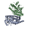
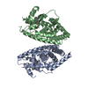
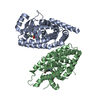
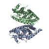
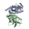
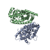
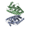
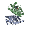
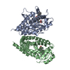
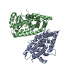
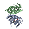
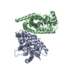
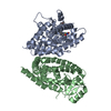
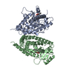
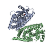
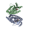
Entry Database : PDB / ID : 3vsoTitle Human PPAR gamma ligand binding domain in complex with a gamma selective agonist mekt21 Peroxisome proliferator-activated receptor gamma Keywords / / Function / homology Function Domain/homology Component
/ / / / / / / / / / / / / / / / / / / / / / / / / / / / / / / / / / / / / / / / / / / / / / / / / / / / / / / / / / / / / / / / / / / / / / / / / / / / / / / / / / / / / / / / / / / / / / / / / / / / / / / / / / / / / / / / / / / / / / / / / / / / / / / / / / / / / / / / / Biological species Homo sapiens (human)Method / / / Resolution : 2 Å Authors Oyama, T. / Waku, T. / Ohashi, M. / Morikawa, K. / Miyachi, H. Journal : Bioorg.Med.Chem. / Year : 2013Title : Design and synthesis of a series of alpha-benzyl phenylpropanoic acid-type peroxisome proliferator-activated receptor (PPAR) gamma partial agonists with improved aqueous solubilityAuthors : Ohashi, M. / Oyama, T. / Putranto, E.W. / Waku, T. / Nobusada, H. / Kataoka, K. / Matsuno, K. / Yashiro, M. / Morikawa, K. / Huh, N.H. / Miyachi, H. History Deposition Apr 30, 2012 Deposition site / Processing site Revision 1.0 May 1, 2013 Provider / Type Revision 1.1 Nov 8, 2023 Group Data collection / Database references ... Data collection / Database references / Derived calculations / Refinement description Category chem_comp_atom / chem_comp_bond ... chem_comp_atom / chem_comp_bond / database_2 / pdbx_initial_refinement_model / struct_ref_seq_dif / struct_site Item _database_2.pdbx_DOI / _database_2.pdbx_database_accession ... _database_2.pdbx_DOI / _database_2.pdbx_database_accession / _struct_ref_seq_dif.details / _struct_site.pdbx_auth_asym_id / _struct_site.pdbx_auth_comp_id / _struct_site.pdbx_auth_seq_id
Show all Show less
 Movie
Movie Controller

 Yorodumi
 Open data
 Basic information
 Components
 Keywords
 Function and homology information
 Homo sapiens (human)
 X-RAY DIFFRACTION /
  Authors
 Citation
 Structure visualization
 Downloads & links
 Other downloads
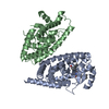


 PDBj
PDBj Assembly
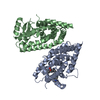


 Mass: 509.596 Da / Num. of mol.: 1 / Source method: obtained synthetically / Formula: C31H31N3O4
 Mass: 18.015 Da / Num. of mol.: 117 / Source method: isolated from a natural source / Formula: H2O
 Sample preparation
 / Beamline: BL38B1 / Wavelength: 1 Å
 Processing