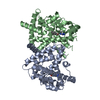
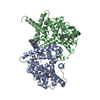
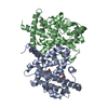
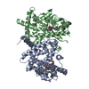
[English] 日本語
Yorodumi
- PDB-5tto: X-ray crystal structure of PPARgamma in complex with SR1643 -

+
Open data


ID or keywords:

Loading...

-
Basic information

Entry
Database: PDB / ID: 5tto
TitleX-ray crystal structure of PPARgamma in complex with SR1643
ComponentsPeroxisome proliferator-activated receptor gamma
KeywordsTRANSCRIPTION / Nuclear Receptor
Function / homology
Function and homology information


prostaglandin receptor activity / regulation of cholesterol transporter activity / negative regulation of connective tissue replacement involved in inflammatory response wound healing / negative regulation of receptor signaling pathway via STAT / MECP2 regulates transcription factors / negative regulation of extracellular matrix assembly / negative regulation of vascular endothelial cell proliferation / negative regulation of cellular response to transforming growth factor beta stimulus / negative regulation of cardiac muscle hypertrophy in response to stress / arachidonic acid binding ...prostaglandin receptor activity / regulation of cholesterol transporter activity / negative regulation of connective tissue replacement involved in inflammatory response wound healing / negative regulation of receptor signaling pathway via STAT / MECP2 regulates transcription factors / negative regulation of extracellular matrix assembly / negative regulation of vascular endothelial cell proliferation / negative regulation of cellular response to transforming growth factor beta stimulus / negative regulation of cardiac muscle hypertrophy in response to stress / arachidonic acid binding / positive regulation of low-density lipoprotein receptor activity / positive regulation of adiponectin secretion / lipoprotein transport / negative regulation of sequestering of triglyceride / DNA binding domain binding / macrophage derived foam cell differentiation / positive regulation of vascular associated smooth muscle cell apoptotic process / STAT family protein binding / positive regulation of fatty acid metabolic process / response to lipid / negative regulation of SMAD protein signal transduction / LBD domain binding / negative regulation of type II interferon-mediated signaling pathway / negative regulation of cholesterol storage / E-box binding / alpha-actinin binding / lipid homeostasis / negative regulation of vascular associated smooth muscle cell proliferation / R-SMAD binding / monocyte differentiation / negative regulation of macrophage derived foam cell differentiation / negative regulation of blood vessel endothelial cell migration / negative regulation of lipid storage / cellular response to low-density lipoprotein particle stimulus / negative regulation of BMP signaling pathway / white fat cell differentiation / negative regulation of mitochondrial fission / positive regulation of cholesterol efflux / retinoic acid receptor signaling pathway / positive regulation of fat cell differentiation / negative regulation of osteoblast differentiation / cell fate commitment / positive regulation of DNA binding / BMP signaling pathway / long-chain fatty acid transport / nuclear retinoid X receptor binding / regulation of cellular response to insulin stimulus / cell maturation / negative regulation of signaling receptor activity / epithelial cell differentiation / positive regulation of adipose tissue development / peroxisome proliferator activated receptor signaling pathway / hormone-mediated signaling pathway / negative regulation of angiogenesis / response to nutrient / negative regulation of miRNA transcription / negative regulation of MAP kinase activity / fatty acid metabolic process / Regulation of PTEN gene transcription / transcription coregulator binding / negative regulation of smooth muscle cell proliferation / peptide binding / negative regulation of transforming growth factor beta receptor signaling pathway / SUMOylation of intracellular receptors / mRNA transcription by RNA polymerase II / placenta development / regulation of circadian rhythm / lipid metabolic process / PPARA activates gene expression / regulation of blood pressure / positive regulation of DNA-binding transcription factor activity / DNA-binding transcription repressor activity, RNA polymerase II-specific / positive regulation of miRNA transcription / negative regulation of inflammatory response / Transcriptional regulation of white adipocyte differentiation / Nuclear Receptor transcription pathway / RNA polymerase II transcription regulator complex / cellular response to insulin stimulus / nuclear receptor activity / activation of cysteine-type endopeptidase activity involved in apoptotic process / : / rhythmic process / glucose homeostasis / cellular response to hypoxia / double-stranded DNA binding / DNA-binding transcription activator activity, RNA polymerase II-specific / DNA-binding transcription factor binding / sequence-specific DNA binding / nucleic acid binding / cell differentiation / receptor complex / transcription cis-regulatory region binding / DNA-binding transcription factor activity, RNA polymerase II-specific / RNA polymerase II cis-regulatory region sequence-specific DNA binding / DNA-binding transcription factor activity / negative regulation of gene expression / intracellular membrane-bounded organelle / innate immune response / negative regulation of DNA-templated transcription / chromatin binding
Similarity search - Function
Peroxisome proliferator-activated receptor gamma / Peroxisome proliferator-activated receptor gamma, N-terminal / PPAR gamma N-terminal region / Peroxisome proliferator-activated receptor / Retinoid X Receptor / Retinoid X Receptor / Nuclear hormone receptor / Nuclear hormones receptors DNA-binding region signature. / Zinc finger, nuclear hormone receptor-type / Zinc finger, C4 type (two domains) ...Peroxisome proliferator-activated receptor gamma / Peroxisome proliferator-activated receptor gamma, N-terminal / PPAR gamma N-terminal region / Peroxisome proliferator-activated receptor / Retinoid X Receptor / Retinoid X Receptor / Nuclear hormone receptor / Nuclear hormones receptors DNA-binding region signature. / Zinc finger, nuclear hormone receptor-type / Zinc finger, C4 type (two domains) / Nuclear hormone receptors DNA-binding domain profile. / c4 zinc finger in nuclear hormone receptors / Nuclear hormone receptor, ligand-binding domain / Nuclear hormone receptor-like domain superfamily / Ligand-binding domain of nuclear hormone receptor / Nuclear receptor (NR) ligand-binding (LBD) domain profile. / Ligand binding domain of hormone receptors / Zinc finger, NHR/GATA-type / Orthogonal Bundle / Mainly Alpha
Similarity search - Domain/homology
Chem-7KK / Peroxisome proliferator-activated receptor gamma
Similarity search - Component
Biological speciesHomo sapiens (human)
MethodX-RAY DIFFRACTION / SYNCHROTRON / MOLECULAR REPLACEMENT / molecular replacement / Resolution: 2.246 Å
AuthorsBruning, J.B. / Frkic, R.L. / Griffin, P. / Kamenecka, T. / Abell, A.
CitationJournal: J. Med. Chem. / Year: 2017
Title: Structure-Activity Relationship of 2,4-Dichloro-N-(3,5-dichloro-4-(quinolin-3-yloxy)phenyl)benzenesulfonamide (INT131) Analogs for PPAR gamma-Targeted Antidiabetics.
Authors: Frkic, R.L. / He, Y. / Rodriguez, B.B. / Chang, M.R. / Kuruvilla, D. / Ciesla, A. / Abell, A.D. / Kamenecka, T.M. / Griffin, P.R. / Bruning, J.B.
History
DepositionNov 4, 2016Deposition site: RCSB / Processing site: RCSB
Revision 1.0May 24, 2017Provider: repository / Type: Initial release
Revision 1.1Jun 21, 2017Group: Database references / Category: citation / citation_author
Item: _citation.country / _citation.journal_id_ASTM ..._citation.country / _citation.journal_id_ASTM / _citation.journal_id_CSD / _citation.journal_volume / _citation.page_first / _citation.page_last / _citation.title
Revision 1.2Oct 4, 2023Group: Data collection / Database references / Refinement description
Category: chem_comp_atom / chem_comp_bond ...chem_comp_atom / chem_comp_bond / database_2 / pdbx_initial_refinement_model
Item: _database_2.pdbx_DOI / _database_2.pdbx_database_accession

-
Structure visualization

Structure viewerMolecule:
MolmilJmol/JSmol

Downloads & links

-
Assembly

Deposited unit
A: Peroxisome proliferator-activated receptor gamma
B: Peroxisome proliferator-activated receptor gamma
hetero molecules


Theoretical massNumber of molelcules
Total (without water)63,5374
Polymers62,4162
Non-polymers1,1202
Water2,864159
1
A: Peroxisome proliferator-activated receptor gamma
hetero molecules

B: Peroxisome proliferator-activated receptor gamma
hetero molecules


Theoretical massNumber of molelcules
Total (without water)63,5374
Polymers62,4162
Non-polymers1,1202
Water362
TypeNameSymmetry operationNumber
identity operation1_555x,y,z1
crystal symmetry operation3_455x-1/2,y+1/2,z1
Buried area2010 Å2
ΔGint-11 kcal/mol
Surface area24040 Å2
MethodPISA
Unit cell
Length a, b, c (Å)93.110, 62.240, 118.880
Angle α, β, γ (deg.)90.000, 102.200, 90.000
Int Tables number5
Space group name H-MC121

-
Components

#1: Protein Peroxisome proliferator-activated receptor gamma / PPAR-gamma / Nuclear receptor subfamily 1 group C member 3


Mass: 31208.236 Da / Num. of mol.: 2 / Fragment: residues 233-505
Source method: isolated from a genetically manipulated source
Source: (gene. exp.) Homo sapiens (human) / Gene: PPARG, NR1C3 / Plasmid: pET11 / Production host: Escherichia coli (E. coli) / Strain (production host): BL21DE3 / References: UniProt: P37231
#2: Chemical ChemComp-7KK / 4-bromo-N-{3,5-dichloro-4-[(quinolin-3-yl)oxy]phenyl}-2,5-difluorobenzene-1-sulfonamide


Mass: 560.195 Da / Num. of mol.: 2 / Source method: obtained synthetically / Formula: C21H11BrCl2F2N2O3S
#3: Water ChemComp-HOH / water


Mass: 18.015 Da / Num. of mol.: 159 / Source method: isolated from a natural source / Formula: H2O

-
Experimental details

-
Experiment

ExperimentMethod: X-RAY DIFFRACTION / Number of used crystals: 1

-
Sample preparation

CrystalDensity Matthews: 2.7 Å3/Da / Density % sol: 54.4 % / Description: cubic
Crystal growTemperature: 289 K / Method: vapor diffusion, hanging drop / pH: 8 / Details: 2M ammonium sulfate

-
Data collection

DiffractionMean temperature: 100 K
Diffraction sourceSource: SYNCHROTRON / Site: APS / Beamline: 22-ID / Wavelength: 1 Å
DetectorType: MARMOSAIC 300 mm CCD / Detector: CCD / Date: Nov 22, 2011
Details: Si 111. Rosenbaum-Rock double-crystal monochromator: liquid nitrogen cooled; sagitally focusing 2nd crystal, Rosenbaum-Rock vertical focusing mirror
RadiationMonochromator: double crystal - liquid nitrogen cooled / Protocol: SINGLE WAVELENGTH / Monochromatic (M) / Laue (L): M / Scattering type: x-ray
Radiation wavelengthWavelength: 1 Å / Relative weight: 1
ReflectionResolution: 2.246→58.1 Å / Num. obs: 31321 / % possible obs: 98 % / Redundancy: 4.7 % / Biso Wilson estimate: 47.98 Å2 / CC1/2: 0.998 / Rmerge(I) obs: 0.053 / Rsym value: 0.053 / Net I/σ(I): 15.3
Reflection shell
Resolution (Å)Redundancy (%)Rmerge(I) obsCC1/2Diffraction-ID% possible all
2.25-2.324.60.2050.973199.7
8.98-58.13.20.0680.986126.9

-
Phasing

PhasingMethod: molecular replacement
Phasing MRModel details: Phaser MODE: MR_AUTO
Highest resolutionLowest resolution
Rotation2.25 Å45.5 Å
Translation2.25 Å45.5 Å

-
Processing

Software
NameVersionClassification
Aimless0.5.25data scaling
PHASER2.6.1phasing
PHENIX(1.10.1_2155)refinement
PDB_EXTRACT3.2data extraction
MOSFLMdata reduction
PHASER2.6.1phasing
RefinementMethod to determine structure: MOLECULAR REPLACEMENT
Starting model: 3FUR
Resolution: 2.246→45.504 Å / SU ML: 0.3 / Cross valid method: THROUGHOUT / σ(F): 1.34 / Phase error: 28.29
RfactorNum. reflection% reflectionSelection details
Rfree0.2555 1536 4.91 %random
Rwork0.2039 ---
obs0.2065 31313 97.96 %-
Solvent computationShrinkage radii: 0.9 Å / VDW probe radii: 1.11 Å
Displacement parametersBiso max: 124.71 Å2 / Biso mean: 61.2923 Å2 / Biso min: 30.81 Å2
Refinement stepCycle: final / Resolution: 2.246→45.504 Å
ProteinNucleic acidLigandSolventTotal
Num. atoms4097 0 64 159 4320
Biso mean--63.2 58.81 -
Num. residues----518
Refine LS restraints
Refine-IDTypeDev idealNumber
X-RAY DIFFRACTIONf_bond_d0.0044288
X-RAY DIFFRACTIONf_angle_d0.6625806
X-RAY DIFFRACTIONf_chiral_restr0.041665
X-RAY DIFFRACTIONf_plane_restr0.004733
X-RAY DIFFRACTIONf_dihedral_angle_d14.9172614
LS refinement shell

Refine-ID: X-RAY DIFFRACTION / Total num. of bins used: 11

Resolution (Å)Rfactor RfreeNum. reflection RfreeRfactor RworkNum. reflection RworkNum. reflection all% reflection obs (%)
2.2461-2.31860.31971380.268927562894100
2.3186-2.40140.36321460.254926962842100
2.4014-2.49760.31991300.241727742904100
2.4976-2.61120.29341500.231427322882100
2.6112-2.74890.27641460.228527402886100
2.7489-2.92110.28991210.23127822903100
2.9211-3.14660.26881280.234927612889100
3.1466-3.46310.2721390.213727772916100
3.4631-3.9640.22411500.184827582908100
3.964-4.99320.23141690.16327432912100
4.9932-45.51290.24621190.20652258237779

+
About Yorodumi

-
News

-
Feb 9, 2022. New format data for meta-information of EMDB entries

New format data for meta-information of EMDB entries

  • Version 3 of the EMDB header file is now the official format.
  • The previous official version 1.9 will be removed from the archive.

Related info.:EMDB header

External links:wwPDB to switch to version 3 of the EMDB data model

-
Aug 12, 2020. Covid-19 info

Covid-19 info

URL: https://pdbjlvh1.pdbj.org/emnavi/covid19.php

New page: Covid-19 featured information page in EM Navigator.

Related info.:Covid-19 info / Mar 5, 2020. Novel coronavirus structure data

+
Mar 5, 2020. Novel coronavirus structure data

Novel coronavirus structure data

Related info.:Yorodumi Speices / Aug 12, 2020. Covid-19 info

External links:COVID-19 featured content - PDBj / Molecule of the Month (242):Coronavirus Proteases

+
Jan 31, 2019. EMDB accession codes are about to change! (news from PDBe EMDB page)

EMDB accession codes are about to change! (news from PDBe EMDB page)

  • The allocation of 4 digits for EMDB accession codes will soon come to an end. Whilst these codes will remain in use, new EMDB accession codes will include an additional digit and will expand incrementally as the available range of codes is exhausted. The current 4-digit format prefixed with “EMD-” (i.e. EMD-XXXX) will advance to a 5-digit format (i.e. EMD-XXXXX), and so on. It is currently estimated that the 4-digit codes will be depleted around Spring 2019, at which point the 5-digit format will come into force.
  • The EM Navigator/Yorodumi systems omit the EMD- prefix.

Related info.:Q: What is EMD? / ID/Accession-code notation in Yorodumi/EM Navigator

External links:EMDB Accession Codes are Changing Soon! / Contact to PDBj

+
Jul 12, 2017. Major update of PDB

Major update of PDB

  • wwPDB released updated PDB data conforming to the new PDBx/mmCIF dictionary.
  • This is a major update changing the version number from 4 to 5, and with Remediation, in which all the entries are updated.
  • In this update, many items about electron microscopy experimental information are reorganized (e.g. em_software).
  • Now, EM Navigator and Yorodumi are based on the updated data.

External links:wwPDB Remediation / Enriched Model Files Conforming to OneDep Data Standards Now Available in the PDB FTP Archive

-
Yorodumi

Thousand views of thousand structures

  • Yorodumi is a browser for structure data from EMDB, PDB, SASBDB, etc.
  • This page is also the successor to EM Navigator detail page, and also detail information page/front-end page for Omokage search.
  • The word "yorodu" (or yorozu) is an old Japanese word meaning "ten thousand". "mi" (miru) is to see.

Related info.:EMDB / PDB / SASBDB / Comparison of 3 databanks / Yorodumi Search / Aug 31, 2016. New EM Navigator & Yorodumi / Yorodumi Papers / Jmol/JSmol / Function and homology information / Changes in new EM Navigator and Yorodumi

Read more