| Entry | Database: PDB / ID: 3x1h
|
|---|
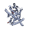
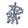
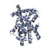
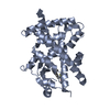
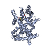
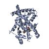
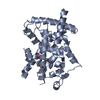
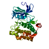
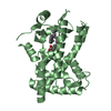
| Title | hPPARgamma Ligand binding domain in complex with 5-oxo-tricosahexaenoic acid |
|---|
 Components Components | Peroxisome proliferator-activated receptor gamma |
|---|
 Keywords Keywords | TRANSCRIPTION / NUCLEAR RECEPTOR / TRANSCRIPTION REGULATION / LIGAND BINDING DOMAIN / DIABETES MELLITUS / ZINC-FINGER / DNA-BINDING / OBESITY |
|---|
| Function / homology |  Function and homology information Function and homology information
prostaglandin receptor activity / negative regulation of receptor signaling pathway via STAT / MECP2 regulates transcription factors / beige fat cell differentiation / negative regulation of vascular endothelial cell proliferation / negative regulation of extracellular matrix assembly / negative regulation of connective tissue replacement involved in inflammatory response wound healing / positive regulation of cholesterol transport / negative regulation of cellular response to transforming growth factor beta stimulus / arachidonate binding ...prostaglandin receptor activity / negative regulation of receptor signaling pathway via STAT / MECP2 regulates transcription factors / beige fat cell differentiation / negative regulation of vascular endothelial cell proliferation / negative regulation of extracellular matrix assembly / negative regulation of connective tissue replacement involved in inflammatory response wound healing / positive regulation of cholesterol transport / negative regulation of cellular response to transforming growth factor beta stimulus / arachidonate binding / positive regulation of adiponectin secretion / DNA binding domain binding / positive regulation of vascular associated smooth muscle cell apoptotic process / negative regulation of cardiac muscle hypertrophy in response to stress / positive regulation of fatty acid metabolic process / positive regulation of lipid metabolic process / STAT family protein binding / WW domain binding / negative regulation of type II interferon-mediated signaling pathway / LBD domain binding / negative regulation of cholesterol storage / response to lipid / positive regulation of lipoprotein transport / negative regulation of SMAD protein signal transduction / lipid homeostasis / E-box binding / R-SMAD binding / negative regulation of blood vessel endothelial cell migration / white fat cell differentiation / monocyte differentiation / negative regulation of vascular associated smooth muscle cell proliferation / negative regulation of BMP signaling pathway / alpha-actinin binding / negative regulation of macrophage derived foam cell differentiation / negative regulation of lipid storage / BMP signaling pathway / positive regulation of cholesterol efflux / cell fate commitment / cellular response to low-density lipoprotein particle stimulus / long-chain fatty acid transport / negative regulation of mitochondrial fission / fat cell differentiation / nuclear retinoid X receptor binding / negative regulation of osteoblast differentiation / positive regulation of fat cell differentiation / retinoic acid receptor signaling pathway / Transcriptional regulation of brown and beige adipocyte differentiation by EBF2 / intracellular receptor signaling pathway / peroxisome proliferator activated receptor signaling pathway / hormone-mediated signaling pathway / cell maturation / negative regulation of MAPK cascade / positive regulation of adipose tissue development / peptide binding / epithelial cell differentiation / regulation of cellular response to insulin stimulus / response to nutrient / negative regulation of miRNA transcription / placenta development / negative regulation of angiogenesis / brown fat cell differentiation / fatty acid metabolic process / positive regulation of apoptotic signaling pathway / Regulation of PTEN gene transcription / transcription coregulator binding / SUMOylation of intracellular receptors / negative regulation of smooth muscle cell proliferation / negative regulation of transforming growth factor beta receptor signaling pathway / PPARA activates gene expression / Nuclear Receptor transcription pathway / Transcriptional regulation of white adipocyte differentiation / regulation of circadian rhythm / negative regulation of inflammatory response / positive regulation of miRNA transcription / DNA-binding transcription repressor activity, RNA polymerase II-specific / mRNA transcription by RNA polymerase II / nuclear receptor activity / regulation of blood pressure / RNA polymerase II transcription regulator complex / cellular response to insulin stimulus / rhythmic process / glucose homeostasis / MLL4 and MLL3 complexes regulate expression of PPARG target genes in adipogenesis and hepatic steatosis / double-stranded DNA binding / DNA-binding transcription activator activity, RNA polymerase II-specific / cellular response to hypoxia / DNA-binding transcription factor binding / sequence-specific DNA binding / nucleic acid binding / DNA-binding transcription factor activity, RNA polymerase II-specific / cell differentiation / signaling receptor complex / transcription cis-regulatory region binding / RNA polymerase II cis-regulatory region sequence-specific DNA binding / DNA-binding transcription factor activity / negative regulation of gene expression / innate immune response / negative regulation of DNA-templated transcription / chromatin binding / positive regulation of gene expressionSimilarity search - Function Peroxisome proliferator-activated receptor gamma / Peroxisome proliferator-activated receptor gamma, N-terminal / PPAR gamma N-terminal region / Peroxisome proliferator-activated receptor / : / Retinoid X Receptor / Retinoid X Receptor / Nuclear hormone receptor / Nuclear hormones receptors DNA-binding region signature. / Zinc finger, nuclear hormone receptor-type ...Peroxisome proliferator-activated receptor gamma / Peroxisome proliferator-activated receptor gamma, N-terminal / PPAR gamma N-terminal region / Peroxisome proliferator-activated receptor / : / Retinoid X Receptor / Retinoid X Receptor / Nuclear hormone receptor / Nuclear hormones receptors DNA-binding region signature. / Zinc finger, nuclear hormone receptor-type / Double treble clef zinc finger, C4 type / Nuclear hormone receptors DNA-binding domain profile. / c4 zinc finger in nuclear hormone receptors / Nuclear hormone receptor, ligand-binding domain / Nuclear hormone receptor-like domain superfamily / Ligand-binding domain of nuclear hormone receptor / Nuclear receptor (NR) ligand-binding (LBD) domain profile. / Ligand binding domain of hormone receptors / Zinc finger, NHR/GATA-type / Orthogonal Bundle / Mainly AlphaSimilarity search - Domain/homology |
|---|
| Biological species |  Homo sapiens (human) Homo sapiens (human) |
|---|
| Method |  X-RAY DIFFRACTION / SYNCHROTRON / MOLECULAR REPLACEMENT / Resolution: 2.3 Å X-RAY DIFFRACTION / SYNCHROTRON / MOLECULAR REPLACEMENT / Resolution: 2.3 Å |
|---|
 Authors Authors | Egawa, D. / Itoh, T. / Yamamoto, K. |
|---|
 Citation Citation | Journal: Bioconjug.Chem. / Year: 2015
Title: Characterization of covalent bond formation between PPAR gamma and oxo-fatty acids.
Authors: Egawa, D. / Itoh, T. / Yamamoto, K. |
|---|
| History | | Deposition | Nov 18, 2014 | Deposition site: PDBJ / Processing site: PDBJ |
|---|
| Revision 1.0 | Apr 8, 2015 | Provider: repository / Type: Initial release |
|---|
| Revision 1.1 | Nov 22, 2017 | Group: Refinement description / Category: software |
|---|
| Revision 1.2 | Aug 24, 2022 | Group: Database references / Derived calculations
Category: citation / database_2 ...citation / database_2 / struct_conn / struct_ref_seq_dif / struct_site
Item: _citation.journal_volume / _citation.page_first ..._citation.journal_volume / _citation.page_first / _citation.page_last / _citation.title / _database_2.pdbx_DOI / _database_2.pdbx_database_accession / _struct_conn.pdbx_dist_value / _struct_conn.pdbx_leaving_atom_flag / _struct_conn.ptnr1_auth_asym_id / _struct_conn.ptnr1_label_asym_id / _struct_conn.ptnr2_auth_asym_id / _struct_conn.ptnr2_label_asym_id / _struct_ref_seq_dif.details / _struct_site.pdbx_auth_asym_id / _struct_site.pdbx_auth_comp_id / _struct_site.pdbx_auth_seq_id |
|---|
| Revision 1.3 | Nov 6, 2024 | Group: Data collection / Structure summary
Category: chem_comp_atom / chem_comp_bond ...chem_comp_atom / chem_comp_bond / pdbx_entry_details / pdbx_modification_feature |
|---|
|
|---|
 Movie
Movie Controller

 Yorodumi
 Open data
 Basic information
 Structure visualization
 Downloads & links
 Other downloads


 PDBj
PDBj Assembly
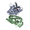


 Mass: 358.514 Da / Num. of mol.: 2 / Source method: obtained synthetically / Formula: C23H34O3
 Mass: 18.015 Da / Num. of mol.: 102 / Source method: isolated from a natural source / Formula: H2O
 Sample preparation
 / Beamline: AR-NW12A / Wavelength: 0.978 Å
 Processing