 Movie
Movie Controller

[English] 日本語
 Yorodumi
- PDB-6dgo: Crystal Structure of Human PPARgamma Ligand Binding Domain in Com... -
+ Open data

- Basic information
| Entry | Database: PDB / ID: 6dgo | ||||||
|---|---|---|---|---|---|---|---|
| Title | Crystal Structure of Human PPARgamma Ligand Binding Domain in Complex with Troglitazone | ||||||
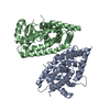
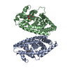
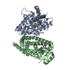
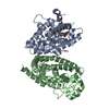
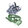
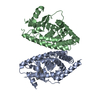
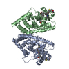
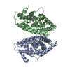
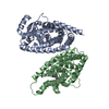
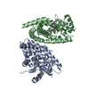
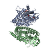
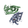
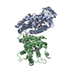
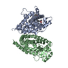
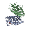
 Components Components | Peroxisome proliferator-activated receptor gamma | ||||||
 Keywords Keywords | transcription/transcription inhibitor / Nuclear receptors / TZDs / Drug design / Therapeutic targets / transcription-transcription inhibitor complex | ||||||
| Function / homology |  Function and homology information Function and homology informationprostaglandin receptor activity / negative regulation of receptor signaling pathway via STAT / MECP2 regulates transcription factors / beige fat cell differentiation / negative regulation of vascular endothelial cell proliferation / negative regulation of extracellular matrix assembly / negative regulation of connective tissue replacement involved in inflammatory response wound healing / positive regulation of cholesterol transport / negative regulation of cellular response to transforming growth factor beta stimulus / arachidonate binding ...prostaglandin receptor activity / negative regulation of receptor signaling pathway via STAT / MECP2 regulates transcription factors / beige fat cell differentiation / negative regulation of vascular endothelial cell proliferation / negative regulation of extracellular matrix assembly / negative regulation of connective tissue replacement involved in inflammatory response wound healing / positive regulation of cholesterol transport / negative regulation of cellular response to transforming growth factor beta stimulus / arachidonate binding / positive regulation of adiponectin secretion / DNA binding domain binding / negative regulation of cardiac muscle hypertrophy in response to stress / positive regulation of vascular associated smooth muscle cell apoptotic process / positive regulation of lipid metabolic process / positive regulation of fatty acid metabolic process / STAT family protein binding / WW domain binding / negative regulation of type II interferon-mediated signaling pathway / LBD domain binding / negative regulation of cholesterol storage / response to lipid / positive regulation of lipoprotein transport / negative regulation of SMAD protein signal transduction / lipid homeostasis / E-box binding / R-SMAD binding / negative regulation of blood vessel endothelial cell migration / white fat cell differentiation / alpha-actinin binding / negative regulation of vascular associated smooth muscle cell proliferation / negative regulation of macrophage derived foam cell differentiation / negative regulation of lipid storage / positive regulation of cholesterol efflux / negative regulation of BMP signaling pathway / monocyte differentiation / cell fate commitment / cellular response to low-density lipoprotein particle stimulus / long-chain fatty acid transport / BMP signaling pathway / negative regulation of mitochondrial fission / negative regulation of osteoblast differentiation / positive regulation of fat cell differentiation / nuclear retinoid X receptor binding / fat cell differentiation / Transcriptional regulation of brown and beige adipocyte differentiation by EBF2 / retinoic acid receptor signaling pathway / intracellular receptor signaling pathway / negative regulation of MAPK cascade / peptide binding / peroxisome proliferator activated receptor signaling pathway / cell maturation / epithelial cell differentiation / hormone-mediated signaling pathway / regulation of cellular response to insulin stimulus / positive regulation of adipose tissue development / response to nutrient / negative regulation of miRNA transcription / brown fat cell differentiation / negative regulation of angiogenesis / placenta development / Regulation of PTEN gene transcription / transcription coregulator binding / SUMOylation of intracellular receptors / positive regulation of apoptotic signaling pathway / negative regulation of smooth muscle cell proliferation / negative regulation of transforming growth factor beta receptor signaling pathway / PPARA activates gene expression / fatty acid metabolic process / Nuclear Receptor transcription pathway / Transcriptional regulation of white adipocyte differentiation / regulation of circadian rhythm / positive regulation of miRNA transcription / mRNA transcription by RNA polymerase II / DNA-binding transcription repressor activity, RNA polymerase II-specific / nuclear receptor activity / negative regulation of inflammatory response / regulation of blood pressure / RNA polymerase II transcription regulator complex / cellular response to insulin stimulus / rhythmic process / glucose homeostasis / MLL4 and MLL3 complexes regulate expression of PPARG target genes in adipogenesis and hepatic steatosis / double-stranded DNA binding / DNA-binding transcription activator activity, RNA polymerase II-specific / cellular response to hypoxia / DNA-binding transcription factor binding / sequence-specific DNA binding / nucleic acid binding / DNA-binding transcription factor activity, RNA polymerase II-specific / cell differentiation / signaling receptor complex / transcription cis-regulatory region binding / RNA polymerase II cis-regulatory region sequence-specific DNA binding / DNA-binding transcription factor activity / negative regulation of gene expression / innate immune response / negative regulation of DNA-templated transcription / chromatin binding / positive regulation of gene expression Similarity search - Function | ||||||
| Biological species |  Homo sapiens (human) Homo sapiens (human) | ||||||
| Method |  X-RAY DIFFRACTION / MOLECULAR REPLACEMENT / Resolution: 3.1 Å X-RAY DIFFRACTION / MOLECULAR REPLACEMENT / Resolution: 3.1 Å | ||||||
 Authors Authors | Shang, J. / Kojetin, D.J. | ||||||
| Funding support |  United States, 1items United States, 1items
| ||||||
 Citation Citation | Journal: Proc.Natl.Acad.Sci.USA / Year: 2019 Title: Quantitative structural assessment of graded receptor agonism. Authors: Shang, J. / Brust, R. / Griffin, P.R. / Kamenecka, T.M. / Kojetin, D.J. | ||||||
| History |
|
- Structure visualization
| Structure viewer | Molecule: MolmilJmol/JSmol |
|---|
- Downloads & links
-Download
| PDBx/mmCIF format | 6dgo.cif.gz | 200.9 KB | Display | PDBx/mmCIF format |
|---|---|---|---|---|
| PDB format | pdb6dgo.ent.gz | 160.5 KB | Display | PDB format |
| PDBx/mmJSON format | 6dgo.json.gz | Tree view | PDBx/mmJSON format | |
| Others |  Other downloads Other downloads |
-Validation report
| Arichive directory | https://data.pdbj.org/pub/pdb/validation_reports/dg/6dgoftp://data.pdbj.org/pub/pdb/validation_reports/dg/6dgo | HTTPS FTP |
|---|
-Related structure data
| Related structure data |  6dglC  6dgqC  6dgrC  6o67C  6o68C 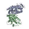 1prgS S: Starting model for refinement C: citing same article ( |
|---|---|
| Similar structure data |


-Links
 PDBj
PDBj- Assembly
| Deposited unit | 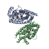
| ||||||||
|---|---|---|---|---|---|---|---|---|---|
| 1 |
| ||||||||
| Unit cell |
|
-Components
| #1: Protein | Mass: 31449.520 Da / Num. of mol.: 2 Source method: isolated from a genetically manipulated source Source: (gene. exp.) Homo sapiens (human) / Gene: PPARG, NR1C3 / Plasmid: pET46 / Production host:  #2: Chemical | ChemComp-GD4 / ( | 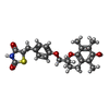  Mass: 441.540 Da / Num. of mol.: 1 / Source method: obtained synthetically / Formula: C24H27NO5S Mass: 441.540 Da / Num. of mol.: 1 / Source method: obtained synthetically / Formula: C24H27NO5S |
|---|
-Experimental details
-Experiment
| Experiment | Method: X-RAY DIFFRACTION / Number of used crystals: 1 |
|---|
- Sample preparation
| Crystal | Density Matthews: 2.45 Å3/Da / Density % sol: 49.76 % |
|---|---|
| Crystal grow | Temperature: 293 K / Method: vapor diffusion, sitting drop / pH: 7.6 / Details: 0.8M SODIUM CITRATE, 100mM MOPS, pH 7.6 |
-Data collection
| Diffraction | Mean temperature: 200 K |
|---|---|
| Diffraction source | Source: ROTATING ANODE / Type: RIGAKU MICROMAX-007 HF / Wavelength: 1.5418 Å |
| Detector | Type: MAR scanner 345 mm plate / Detector: IMAGE PLATE / Date: Apr 19, 2017 |
| Radiation | Protocol: SINGLE WAVELENGTH / Monochromatic (M) / Laue (L): M / Scattering type: x-ray |
| Radiation wavelength | Wavelength: 1.5418 Å / Relative weight: 1 |
| Reflection | Resolution: 3.1→45.374 Å / Num. obs: 12003 / % possible obs: 98.55 % / Redundancy: 1.8 % / Net I/σ(I): 8.48 |
| Reflection shell | Resolution: 3.1→3.211 Å / Redundancy: 1.8 % / Mean I/σ(I) obs: 2.2 / Num. unique obs: 1150 / % possible all: 97.71 |
- Processing
| Software |
| ||||||||||||||||||||||||||||||||||||||||||||||||||||||||||||||||||||||
|---|---|---|---|---|---|---|---|---|---|---|---|---|---|---|---|---|---|---|---|---|---|---|---|---|---|---|---|---|---|---|---|---|---|---|---|---|---|---|---|---|---|---|---|---|---|---|---|---|---|---|---|---|---|---|---|---|---|---|---|---|---|---|---|---|---|---|---|---|---|---|---|
| Refinement | Method to determine structure: MOLECULAR REPLACEMENT Starting model: 1PRG Resolution: 3.1→45.374 Å / SU ML: 0.46 / Cross valid method: THROUGHOUT / σ(F): 1.38 / Phase error: 27.36 / Stereochemistry target values: ML
| ||||||||||||||||||||||||||||||||||||||||||||||||||||||||||||||||||||||
| Solvent computation | Shrinkage radii: 0.9 Å / VDW probe radii: 1.11 Å / Solvent model: FLAT BULK SOLVENT MODEL | ||||||||||||||||||||||||||||||||||||||||||||||||||||||||||||||||||||||
| Displacement parameters | Biso max: 114.8 Å2 / Biso mean: 25.1537 Å2 / Biso min: 2.71 Å2 | ||||||||||||||||||||||||||||||||||||||||||||||||||||||||||||||||||||||
| Refinement step | Cycle: final / Resolution: 3.1→45.374 Å
| ||||||||||||||||||||||||||||||||||||||||||||||||||||||||||||||||||||||
| Refine LS restraints |
| ||||||||||||||||||||||||||||||||||||||||||||||||||||||||||||||||||||||
| LS refinement shell | Refine-ID: X-RAY DIFFRACTION / Rfactor Rfree error: 0 / Total num. of bins used: 9
| ||||||||||||||||||||||||||||||||||||||||||||||||||||||||||||||||||||||
| Refinement TLS params. | Method: refined / Origin x: 21.6425 Å / Origin y: 45.111 Å / Origin z: 25.3652 Å
| ||||||||||||||||||||||||||||||||||||||||||||||||||||||||||||||||||||||
| Refinement TLS group |
|
