 Movie
Movie Controller

[English] 日本語
 Yorodumi
- PDB-3fsu: Crystal Structure of Escherichia coli Methylenetetrahydrofolate R... -
+ Open data

- Basic information
| Entry | Database: PDB / ID: 3fsu | ||||||
|---|---|---|---|---|---|---|---|
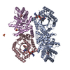
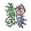
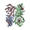
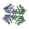
| Title | Crystal Structure of Escherichia coli Methylenetetrahydrofolate Reductase Double Mutant Phe223LeuGlu28Gln complexed with methyltetrahydrofolate | ||||||
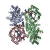
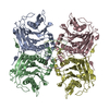
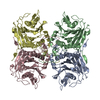
 Components Components | 5,10-methylenetetrahydrofolate reductase | ||||||
 Keywords Keywords | OXIDOREDUCTASE / TIM BARREL / FLAVIN / REDUCTASE / METHYLTETRAHYDROFOLATE / Amino-acid biosynthesis / FAD / Flavoprotein / Methionine biosynthesis / NAD / NADP | ||||||
| Function / homology |  Function and homology information Function and homology informationmethylenetetrahydrofolate reductase (NADH) / methylenetetrahydrofolate reductase (NADH) activity / methylenetetrahydrofolate reductase [NAD(P)H] activity / : / tetrahydrofolate interconversion / FAD binding / protein-folding chaperone binding / protein-containing complex / cytosol Similarity search - Function | ||||||
| Biological species |  | ||||||
| Method |  X-RAY DIFFRACTION / SYNCHROTRON / Resolution: 1.7 Å X-RAY DIFFRACTION / SYNCHROTRON / Resolution: 1.7 Å | ||||||
 Authors Authors | Tanner, J.J. | ||||||
 Citation Citation | Journal: Biochemistry / Year: 2009 Title: Functional role for the conformationally mobile phenylalanine 223 in the reaction of methylenetetrahydrofolate reductase from Escherichia coli. Authors: Lee, M.N. / Takawira, D. / Nikolova, A.P. / Ballou, D.P. / Furtado, V.C. / Phung, N.L. / Still, B.R. / Thorstad, M.K. / Tanner, J.J. / Trimmer, E.E. | ||||||
| History |
|
- Structure visualization
| Structure viewer | Molecule: MolmilJmol/JSmol |
|---|
- Downloads & links
-Download
| PDBx/mmCIF format | 3fsu.cif.gz | 345.6 KB | Display | PDBx/mmCIF format |
|---|---|---|---|---|
| PDB format | pdb3fsu.ent.gz | 280.2 KB | Display | PDB format |
| PDBx/mmJSON format | 3fsu.json.gz | Tree view | PDBx/mmJSON format | |
| Others |  Other downloads Other downloads |
-Validation report
| Arichive directory | https://data.pdbj.org/pub/pdb/validation_reports/fs/3fsuftp://data.pdbj.org/pub/pdb/validation_reports/fs/3fsu | HTTPS FTP |
|---|
-Related structure data
| Related structure data |  3fstC  1zp4S C: citing same article ( S: Starting model for refinement |
|---|---|
| Similar structure data |


-Links
 PDBj
PDBj- Assembly
| Deposited unit | 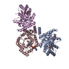
| ||||||||
|---|---|---|---|---|---|---|---|---|---|
| 1 | 
| ||||||||
| Unit cell |
| ||||||||
| Components on special symmetry positions |
|
-Components
-Protein , 1 types, 3 molecules ACE
| #1: Protein | Mass: 34180.852 Da / Num. of mol.: 3 / Mutation: F223L,E28Q Source method: isolated from a genetically manipulated source Source: (gene. exp.) References: UniProt: P0AEZ1, methylenetetrahydrofolate reductase [NAD(P)H] |
|---|
-Non-polymers , 5 types, 407 molecules 
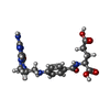
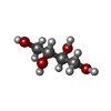


| #2: Chemical |  Mass: 785.550 Da / Num. of mol.: 3 / Source method: obtained synthetically / Formula: C27H33N9O15P2 / Comment: FAD*YM Mass: 785.550 Da / Num. of mol.: 3 / Source method: obtained synthetically / Formula: C27H33N9O15P2 / Comment: FAD*YM#3: Chemical |  Mass: 459.456 Da / Num. of mol.: 2 / Source method: obtained synthetically / Formula: C20H25N7O6 Mass: 459.456 Da / Num. of mol.: 2 / Source method: obtained synthetically / Formula: C20H25N7O6#4: Chemical | ChemComp-MRY / |  Mass: 122.120 Da / Num. of mol.: 1 / Source method: obtained synthetically / Formula: C4H10O4 Mass: 122.120 Da / Num. of mol.: 1 / Source method: obtained synthetically / Formula: C4H10O4#5: Chemical | ChemComp-SO4 / |  Mass: 96.063 Da / Num. of mol.: 1 / Source method: obtained synthetically / Formula: SO4 Mass: 96.063 Da / Num. of mol.: 1 / Source method: obtained synthetically / Formula: SO4#6: Water | ChemComp-HOH / | Mass: 18.015 Da / Num. of mol.: 400 / Source method: isolated from a natural source / Formula: H2O |
|---|
-Experimental details
-Experiment
| Experiment | Method: X-RAY DIFFRACTION / Number of used crystals: 1 |
|---|
- Sample preparation
| Crystal | Density Matthews: 2.66 Å3/Da / Density % sol: 53.83 % |
|---|---|
| Crystal grow | Temperature: 298 K / Method: vapor diffusion / pH: 5 Details: 100 mM sodium cacodylate buffer pH 5.0- 5.5, 225 mM Li2SO4, 5 % ethanol and 10 - 12 % PEG 4000, vapor diffusion, temperature 298K |
-Data collection
| Diffraction | Mean temperature: 100 K | ||||||||||||||||||||||||||||||||||||||||||||||||||||||||||||||||||||||||||||||||||||||||
|---|---|---|---|---|---|---|---|---|---|---|---|---|---|---|---|---|---|---|---|---|---|---|---|---|---|---|---|---|---|---|---|---|---|---|---|---|---|---|---|---|---|---|---|---|---|---|---|---|---|---|---|---|---|---|---|---|---|---|---|---|---|---|---|---|---|---|---|---|---|---|---|---|---|---|---|---|---|---|---|---|---|---|---|---|---|---|---|---|---|
| Diffraction source | Source: SYNCHROTRON / Site: ALS  / Beamline: 4.2.2 / Wavelength: 1 Å / Beamline: 4.2.2 / Wavelength: 1 Å | ||||||||||||||||||||||||||||||||||||||||||||||||||||||||||||||||||||||||||||||||||||||||
| Detector | Type: NOIR-1 / Detector: CCD / Date: Jun 1, 2008 | ||||||||||||||||||||||||||||||||||||||||||||||||||||||||||||||||||||||||||||||||||||||||
| Radiation | Monochromator: beamline 4.2.2 optics / Protocol: SINGLE WAVELENGTH / Monochromatic (M) / Laue (L): M / Scattering type: x-ray | ||||||||||||||||||||||||||||||||||||||||||||||||||||||||||||||||||||||||||||||||||||||||
| Radiation wavelength | Wavelength: 1 Å / Relative weight: 1 | ||||||||||||||||||||||||||||||||||||||||||||||||||||||||||||||||||||||||||||||||||||||||
| Reflection | Resolution: 1.7→43.71 Å / Num. obs: 117437 / % possible obs: 99.8 % / Redundancy: 3.75 % / Rmerge(I) obs: 0.033 / Χ2: 0.96 / Scaling rejects: 3332 | ||||||||||||||||||||||||||||||||||||||||||||||||||||||||||||||||||||||||||||||||||||||||
| Reflection shell |
|
- Processing
| Software |
| ||||||||||||||||||||||||||||||||||||||||||||||||||||||||||||||||||||||||||||||||||||||||||||||||||||
|---|---|---|---|---|---|---|---|---|---|---|---|---|---|---|---|---|---|---|---|---|---|---|---|---|---|---|---|---|---|---|---|---|---|---|---|---|---|---|---|---|---|---|---|---|---|---|---|---|---|---|---|---|---|---|---|---|---|---|---|---|---|---|---|---|---|---|---|---|---|---|---|---|---|---|---|---|---|---|---|---|---|---|---|---|---|---|---|---|---|---|---|---|---|---|---|---|---|---|---|---|---|
| Refinement | Starting model: PDB ENTRY 1zp4 Resolution: 1.7→43.707 Å / Occupancy max: 1 / Occupancy min: 0.5 / FOM work R set: 0.851 / SU ML: 0.25 / Isotropic thermal model: isotropic / Cross valid method: THROUGHOUT / σ(F): 1.34 / Stereochemistry target values: ML
| ||||||||||||||||||||||||||||||||||||||||||||||||||||||||||||||||||||||||||||||||||||||||||||||||||||
| Solvent computation | Shrinkage radii: 0.9 Å / VDW probe radii: 1.11 Å / Solvent model: FLAT BULK SOLVENT MODEL / Bsol: 45.514 Å2 / ksol: 0.37 e/Å3 | ||||||||||||||||||||||||||||||||||||||||||||||||||||||||||||||||||||||||||||||||||||||||||||||||||||
| Displacement parameters | Biso max: 253.76 Å2 / Biso mean: 31.971 Å2 / Biso min: 11.17 Å2
| ||||||||||||||||||||||||||||||||||||||||||||||||||||||||||||||||||||||||||||||||||||||||||||||||||||
| Refinement step | Cycle: LAST / Resolution: 1.7→43.707 Å
| ||||||||||||||||||||||||||||||||||||||||||||||||||||||||||||||||||||||||||||||||||||||||||||||||||||
| Refine LS restraints |
| ||||||||||||||||||||||||||||||||||||||||||||||||||||||||||||||||||||||||||||||||||||||||||||||||||||
| LS refinement shell | Refine-ID: X-RAY DIFFRACTION / Total num. of bins used: 10
| ||||||||||||||||||||||||||||||||||||||||||||||||||||||||||||||||||||||||||||||||||||||||||||||||||||
| Refinement TLS params. | Method: refined / Refine-ID: X-RAY DIFFRACTION
| ||||||||||||||||||||||||||||||||||||||||||||||||||||||||||||||||||||||||||||||||||||||||||||||||||||
| Refinement TLS group |
|
