 Movie
Movie Controller

[English] 日本語
 Yorodumi
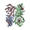
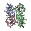
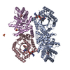
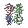
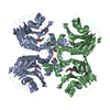
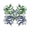
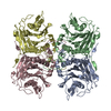
- PDB-2fmn: Ala177Val mutant of E. coli Methylenetetrahydrofolate Reductase c... -
+ Open data

- Basic information
| Entry | Database: PDB / ID: 2fmn | ||||||
|---|---|---|---|---|---|---|---|
| Title | Ala177Val mutant of E. coli Methylenetetrahydrofolate Reductase complex with LY309887 | ||||||
 Components Components | 5,10-methylenetetrahydrofolate reductase | ||||||
 Keywords Keywords | OXIDOREDUCTASE / TIM BARREL / FLAVIN / REDUCTASE | ||||||
| Function / homology |  Function and homology information Function and homology informationmethylenetetrahydrofolate reductase (NADH) / methylenetetrahydrofolate reductase (NADH) activity / methylenetetrahydrofolate reductase [NAD(P)H] activity / : / tetrahydrofolate interconversion / FAD binding / protein-folding chaperone binding / protein-containing complex / cytosol Similarity search - Function | ||||||
| Biological species |  | ||||||
| Method |  X-RAY DIFFRACTION / MOLECULAR REPLACEMENT / Resolution: 2.05 Å X-RAY DIFFRACTION / MOLECULAR REPLACEMENT / Resolution: 2.05 Å | ||||||
 Authors Authors | Pejchal, R. / Campbell, E. / Guenther, B.D. / Lennon, B.W. / Matthews, R.G. / Ludwig, M.L. | ||||||
 Citation Citation | Journal: Biochemistry / Year: 2006 Title: Structural Perturbations in the Ala -> Val Polymorphism of Methylenetetrahydrofolate Reductase: How Binding of Folates May Protect against Inactivation Authors: Pejchal, R. / Campbell, E. / Guenther, B.D. / Lennon, B.W. / Matthews, R.G. / Ludwig, M.L. | ||||||
| History |
|
- Structure visualization
| Structure viewer | Molecule: MolmilJmol/JSmol |
|---|
- Downloads & links
-Download
| PDBx/mmCIF format | 2fmn.cif.gz | 186.2 KB | Display | PDBx/mmCIF format |
|---|---|---|---|---|
| PDB format | pdb2fmn.ent.gz | 147.2 KB | Display | PDB format |
| PDBx/mmJSON format | 2fmn.json.gz | Tree view | PDBx/mmJSON format | |
| Others |  Other downloads Other downloads |
-Validation report
| Arichive directory | https://data.pdbj.org/pub/pdb/validation_reports/fm/2fmnftp://data.pdbj.org/pub/pdb/validation_reports/fm/2fmn | HTTPS FTP |
|---|
-Related structure data
| Related structure data |  2fmoC  1b5tS C: citing same article ( S: Starting model for refinement |
|---|---|
| Similar structure data |

-Links
 PDBj
PDBj- Assembly
| Deposited unit | 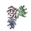
| ||||||||
|---|---|---|---|---|---|---|---|---|---|
| 1 | 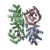
| ||||||||
| Unit cell |
| ||||||||
| Details | This entry contains the crystallographic asymmetric unit, which consists of 3 chains. The second part of the biological assembly contains a symmetry related chain B generated by the two fold axis: -x, y, -z. The biological assembly consists of chains A, B, B', and C, where B' is the symmetry related chain B. |
-Components
| #1: Protein | Mass: 34243.902 Da / Num. of mol.: 3 / Mutation: A177V Source method: isolated from a genetically manipulated source Source: (gene. exp.) References: UniProt: P0AEZ1, methylenetetrahydrofolate reductase [NAD(P)H] #2: Chemical |   Mass: 785.550 Da / Num. of mol.: 3 / Source method: obtained synthetically / Formula: C27H33N9O15P2 / Comment: FAD*YM Mass: 785.550 Da / Num. of mol.: 3 / Source method: obtained synthetically / Formula: C27H33N9O15P2 / Comment: FAD*YM#3: Chemical | 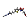  Mass: 449.481 Da / Num. of mol.: 3 / Source method: obtained synthetically / Formula: C19H23N5O6S Mass: 449.481 Da / Num. of mol.: 3 / Source method: obtained synthetically / Formula: C19H23N5O6S#4: Water | ChemComp-HOH / |  Mass: 18.015 Da / Num. of mol.: 281 / Source method: isolated from a natural source / Formula: H2O Mass: 18.015 Da / Num. of mol.: 281 / Source method: isolated from a natural source / Formula: H2O |
|---|
-Experimental details
-Experiment
| Experiment | Method: X-RAY DIFFRACTION / Number of used crystals: 1 |
|---|
- Sample preparation
| Crystal | Density Matthews: 2.64 Å3/Da / Density % sol: 53.39 % |
|---|---|
| Crystal grow | Temperature: 295 K / pH: 6.5 Details: PEG 8000, MAGNESIUM ACETATE, SODIUM CACODYLATE, GLYCEROL, METHYLPENTANEDIOL, LY309887, VAPOR DIFFUSION, HANGING DROP, temperature 295K, pH 6.50 |
-Data collection
| Diffraction | Mean temperature: 100 K |
|---|---|
| Diffraction source | Source: ROTATING ANODE / Type: RIGAKU / Wavelength: 1.5418 |
| Detector | Type: RIGAKU RAXIS IV / Detector: IMAGE PLATE / Date: Jan 1, 2000 |
| Radiation | Protocol: SINGLE WAVELENGTH / Monochromatic (M) / Laue (L): M / Scattering type: x-ray |
| Radiation wavelength | Wavelength: 1.5418 Å / Relative weight: 1 |
| Reflection | Resolution: 2.05→35.8 Å / Num. obs: 62268 / % possible obs: 93.3 % / Observed criterion σ(I): 0 / Biso Wilson estimate: 22.5 Å2 |
| Reflection shell | Resolution: 2.05→2.18 Å / % possible all: 85.1 |
- Processing
| Software |
| ||||||||||||||||||||||||||||||||||||||||||||||||||||||||||||||||||||||||||||||||
|---|---|---|---|---|---|---|---|---|---|---|---|---|---|---|---|---|---|---|---|---|---|---|---|---|---|---|---|---|---|---|---|---|---|---|---|---|---|---|---|---|---|---|---|---|---|---|---|---|---|---|---|---|---|---|---|---|---|---|---|---|---|---|---|---|---|---|---|---|---|---|---|---|---|---|---|---|---|---|---|---|---|
| Refinement | Method to determine structure: MOLECULAR REPLACEMENT Starting model: pdb entry 1B5T Resolution: 2.05→35.78 Å / Rfactor Rfree error: 0.004 / Data cutoff high absF: 547514.57 / Data cutoff low absF: 0 / Isotropic thermal model: RESTRAINED / Cross valid method: THROUGHOUT / σ(F): 0 / Stereochemistry target values: ENGH & HUBER
| ||||||||||||||||||||||||||||||||||||||||||||||||||||||||||||||||||||||||||||||||
| Solvent computation | Solvent model: FLAT MODEL / Bsol: 42.29 Å2 / ksol: 0.34 e/Å3 | ||||||||||||||||||||||||||||||||||||||||||||||||||||||||||||||||||||||||||||||||
| Displacement parameters | Biso mean: 34.2 Å2
| ||||||||||||||||||||||||||||||||||||||||||||||||||||||||||||||||||||||||||||||||
| Refine analyze |
| ||||||||||||||||||||||||||||||||||||||||||||||||||||||||||||||||||||||||||||||||
| Refinement step | Cycle: LAST / Resolution: 2.05→35.78 Å
| ||||||||||||||||||||||||||||||||||||||||||||||||||||||||||||||||||||||||||||||||
| Refine LS restraints |
| ||||||||||||||||||||||||||||||||||||||||||||||||||||||||||||||||||||||||||||||||
| LS refinement shell | Resolution: 2.05→2.18 Å / Rfactor Rfree error: 0.013 / Total num. of bins used: 6
| ||||||||||||||||||||||||||||||||||||||||||||||||||||||||||||||||||||||||||||||||
| Xplor file |
|
