 Movie
Movie Controller

[English] 日本語
 Yorodumi
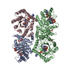
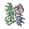
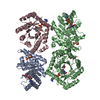
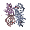
- PDB-1zp4: Glu28Gln mutant of E. coli Methylenetetrahydrofolate Reductase (o... -
+ Open data

- Basic information
| Entry | Database: PDB / ID: 1zp4 | ||||||
|---|---|---|---|---|---|---|---|
| Title | Glu28Gln mutant of E. coli Methylenetetrahydrofolate Reductase (oxidized) complex with Methyltetrahydrofolate | ||||||
 Components Components | 5,10-methylenetetrahydrofolate reductase | ||||||
 Keywords Keywords | OXIDOREDUCTASE / TIM BARREL / METHYLTETRAHYDROFOLATE / FLAVIN / REDUCTASE | ||||||
| Function / homology |  Function and homology information Function and homology informationmethylenetetrahydrofolate reductase [NAD(P)H] / methylenetetrahydrofolate reductase (NADPH) activity / methylenetetrahydrofolate reductase (NADH) / methylenetetrahydrofolate reductase (NADH) activity / methylenetetrahydrofolate reductase [NAD(P)H] activity / : / tetrahydrofolate interconversion / FAD binding / protein-folding chaperone binding / protein-containing complex / cytosol Similarity search - Function | ||||||
| Biological species |  | ||||||
| Method |  X-RAY DIFFRACTION / SYNCHROTRON / MOLECULAR REPLACEMENT / Resolution: 1.85 Å X-RAY DIFFRACTION / SYNCHROTRON / MOLECULAR REPLACEMENT / Resolution: 1.85 Å | ||||||
 Authors Authors | Pejchal, R. / Sargeant, R. / Ludwig, M.L. | ||||||
 Citation Citation | Journal: Biochemistry / Year: 2005 Title: Structures of NADH and CH(3)-H(4)Folate Complexes of Escherichia coli Methylenetetrahydrofolate Reductase Reveal a Spartan Strategy for a Ping-Pong Reaction Authors: Pejchal, R. / Sargeant, R. / Ludwig, M.L. | ||||||
| History |
|
- Structure visualization
| Structure viewer | Molecule: MolmilJmol/JSmol |
|---|
- Downloads & links
-Download
| PDBx/mmCIF format | 1zp4.cif.gz | 191.3 KB | Display | PDBx/mmCIF format |
|---|---|---|---|---|
| PDB format | pdb1zp4.ent.gz | 150.2 KB | Display | PDB format |
| PDBx/mmJSON format | 1zp4.json.gz | Tree view | PDBx/mmJSON format | |
| Others |  Other downloads Other downloads |
-Validation report
| Arichive directory | https://data.pdbj.org/pub/pdb/validation_reports/zp/1zp4ftp://data.pdbj.org/pub/pdb/validation_reports/zp/1zp4 | HTTPS FTP |
|---|
-Related structure data
| Related structure data |  1zp3C  1zptC  1zrqC  1b5tS S: Starting model for refinement C: citing same article ( |
|---|---|
| Similar structure data |

-Links
 PDBj
PDBj- Assembly
| Deposited unit | 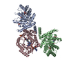
| ||||||||
|---|---|---|---|---|---|---|---|---|---|
| 1 | 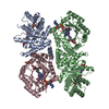
| ||||||||
| Unit cell |
| ||||||||
| Components on special symmetry positions |
| ||||||||
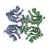
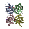
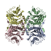
| Details | The biological assembly is a tetramer. It can be generated from the trimer in the asymmetric unit (A,B,C) by the operations: -y, x, -z. A unique tetramer (A,B,B',C' or A',B',B,C) can be picked from the resulting hexamer (A,B,C,A',B',C'). |
-Components
| #1: Protein | Mass: 34214.867 Da / Num. of mol.: 3 / Mutation: E28Q Source method: isolated from a genetically manipulated source Source: (gene. exp.) References: UniProt: P00394, UniProt: P0AEZ1*PLUS, EC: 1.7.99.5 #2: Chemical |   Mass: 96.063 Da / Num. of mol.: 2 / Source method: obtained synthetically / Formula: SO4 Mass: 96.063 Da / Num. of mol.: 2 / Source method: obtained synthetically / Formula: SO4#3: Chemical | 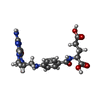  Mass: 459.456 Da / Num. of mol.: 3 / Source method: obtained synthetically / Formula: C20H25N7O6 Mass: 459.456 Da / Num. of mol.: 3 / Source method: obtained synthetically / Formula: C20H25N7O6#4: Chemical |   Mass: 785.550 Da / Num. of mol.: 3 / Source method: obtained synthetically / Formula: C27H33N9O15P2 / Comment: FAD*YM Mass: 785.550 Da / Num. of mol.: 3 / Source method: obtained synthetically / Formula: C27H33N9O15P2 / Comment: FAD*YM#5: Water | ChemComp-HOH / |  Mass: 18.015 Da / Num. of mol.: 509 / Source method: isolated from a natural source / Formula: H2O Mass: 18.015 Da / Num. of mol.: 509 / Source method: isolated from a natural source / Formula: H2O |
|---|
-Experimental details
-Experiment
| Experiment | Method: X-RAY DIFFRACTION / Number of used crystals: 1 |
|---|
- Sample preparation
| Crystal | Density Matthews: 2.69 Å3/Da / Density % sol: 54.2 % |
|---|---|
| Crystal grow | Temperature: 295 K / Method: vapor diffusion, hanging drop / pH: 7.4 Details: PEG 4000, LITHIUM SULFATE, SODIUM CACODYLATE, ETHANOL, MESO-ERYTHRITOL, pH 7.4, VAPOR DIFFUSION, HANGING DROP, temperature 295.0K |
-Data collection
| Diffraction | Mean temperature: 100 K |
|---|---|
| Diffraction source | Source: SYNCHROTRON / Site: APS  / Beamline: 5ID-B / Wavelength: 0.97623 Å / Beamline: 5ID-B / Wavelength: 0.97623 Å |
| Detector | Type: MARMOSAIC 225 mm CCD / Detector: CCD / Date: Mar 10, 2003 |
| Radiation | Protocol: SINGLE WAVELENGTH / Monochromatic (M) / Laue (L): M / Scattering type: x-ray |
| Radiation wavelength | Wavelength: 0.97623 Å / Relative weight: 1 |
| Reflection | Resolution: 1.85→17.27 Å / Num. all: 91950 / Num. obs: 91462 / % possible obs: 99.5 % / Observed criterion σ(F): 0 / Observed criterion σ(I): 0 / Redundancy: 3.6 % / Biso Wilson estimate: 20.1 Å2 / Rsym value: 0.06 / Net I/σ(I): 14.41 |
| Reflection shell | Resolution: 1.85→1.91 Å / Redundancy: 3.65 % / Mean I/σ(I) obs: 3.8 / Rsym value: 0.347 / % possible all: 99.7 |
- Processing
| Software |
| ||||||||||||||||||||||||||||||||||||||||||||||||||||||||||||||||||||||||||||||||
|---|---|---|---|---|---|---|---|---|---|---|---|---|---|---|---|---|---|---|---|---|---|---|---|---|---|---|---|---|---|---|---|---|---|---|---|---|---|---|---|---|---|---|---|---|---|---|---|---|---|---|---|---|---|---|---|---|---|---|---|---|---|---|---|---|---|---|---|---|---|---|---|---|---|---|---|---|---|---|---|---|---|
| Refinement | Method to determine structure: MOLECULAR REPLACEMENT Starting model: PDB ENTRY 1B5T Resolution: 1.85→17.27 Å / Rfactor Rfree error: 0.003 / Data cutoff high absF: 4072347.7 / Data cutoff low absF: 0 / Isotropic thermal model: RESTRAINED / Cross valid method: THROUGHOUT / σ(F): 0 / Stereochemistry target values: Engh & Huber
| ||||||||||||||||||||||||||||||||||||||||||||||||||||||||||||||||||||||||||||||||
| Solvent computation | Solvent model: FLAT MODEL / Bsol: 83.7331 Å2 / ksol: 0.575832 e/Å3 | ||||||||||||||||||||||||||||||||||||||||||||||||||||||||||||||||||||||||||||||||
| Displacement parameters | Biso mean: 28.3 Å2
| ||||||||||||||||||||||||||||||||||||||||||||||||||||||||||||||||||||||||||||||||
| Refine analyze |
| ||||||||||||||||||||||||||||||||||||||||||||||||||||||||||||||||||||||||||||||||
| Refinement step | Cycle: LAST / Resolution: 1.85→17.27 Å
| ||||||||||||||||||||||||||||||||||||||||||||||||||||||||||||||||||||||||||||||||
| Refine LS restraints |
| ||||||||||||||||||||||||||||||||||||||||||||||||||||||||||||||||||||||||||||||||
| LS refinement shell | Resolution: 1.85→1.97 Å / Rfactor Rfree error: 0.011 / Total num. of bins used: 6
| ||||||||||||||||||||||||||||||||||||||||||||||||||||||||||||||||||||||||||||||||
| Xplor file |
|
