 ムービー
ムービー コントローラー


+ データを開く

- 基本情報
基本情報
| 登録情報 | データベース: PDB / ID: 1zp3 | ||||||
|---|---|---|---|---|---|---|---|
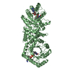
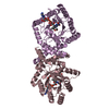
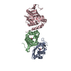
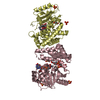
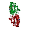
| タイトル | E. coli Methylenetetrahydrofolate Reductase (oxidized) | ||||||
 要素 要素 | 5,10-methylenetetrahydrofolate reductase | ||||||
 キーワード キーワード | OXIDOREDUCTASE / TIM BARREL / FLAVIN / REDUCTASE | ||||||
| 機能・相同性 |  機能・相同性情報 機能・相同性情報methylenetetrahydrofolate reductase [NAD(P)H] / methylenetetrahydrofolate reductase (NADPH) activity / methylenetetrahydrofolate reductase (NADH) / methylenetetrahydrofolate reductase (NADH) activity / methylenetetrahydrofolate reductase [NAD(P)H] activity / : / tetrahydrofolate interconversion / FAD binding / protein-folding chaperone binding / protein-containing complex / cytosol 類似検索 - 分子機能 | ||||||
| 生物種 |  | ||||||
| 手法 |  X線回折 / 分子置換 / 解像度: 1.85 Å X線回折 / 分子置換 / 解像度: 1.85 Å | ||||||
 データ登録者 データ登録者 | Pejchal, R. / Sargeant, R. / Ludwig, M.L. | ||||||
 引用 引用 | ジャーナル: Biochemistry / 年: 2005 タイトル: Structures of NADH and CH(3)-H(4)Folate Complexes of Escherichia coli Methylenetetrahydrofolate Reductase Reveal a Spartan Strategy for a Ping-Pong Reaction 著者: Pejchal, R. / Sargeant, R. / Ludwig, M.L. | ||||||
| 履歴 |
|
- 構造の表示
構造の表示
| 構造ビューア | 分子: MolmilJmol/JSmol |
|---|
- ダウンロードとリンク
-ダウンロード
| PDBx/mmCIF形式 | 1zp3.cif.gz | 187.2 KB | 表示 | PDBx/mmCIF形式 |
|---|---|---|---|---|
| PDB形式 | pdb1zp3.ent.gz | 146.5 KB | 表示 | PDB形式 |
| PDBx/mmJSON形式 | 1zp3.json.gz | ツリー表示 | PDBx/mmJSON形式 | |
| その他 |  その他のダウンロード その他のダウンロード |
-検証レポート
| アーカイブディレクトリ | https://data.pdbj.org/pub/pdb/validation_reports/zp/1zp3ftp://data.pdbj.org/pub/pdb/validation_reports/zp/1zp3 | HTTPS FTP |
|---|
-関連構造データ


-リンク
 PDBj
PDBj- 集合体
集合体
| 登録構造単位 | 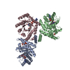
| ||||||||||||
|---|---|---|---|---|---|---|---|---|---|---|---|---|---|
| 1 | 
| ||||||||||||
| 2 | 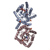
| ||||||||||||
| 3 | 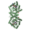
| ||||||||||||
| 単位格子 |
| ||||||||||||
| Components on special symmetry positions |
| ||||||||||||
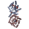
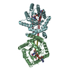
| 詳細 | The biological assembly is a tetramer. It can be generated from the trimer in the asymmetric unit (A,B,C) by the operations: -y, x, -z. A unique tetramer (A,B,B',C' or A',B',B,C) can be picked from the resulting hexamer (A,B,C,A',B',C'). |
-要素
| #1: タンパク質 | 分子量: 34215.852 Da / 分子数: 3 / 由来タイプ: 組換発現 / 由来: (組換発現) #2: 化合物 | ChemComp-SO4 /   分子量: 96.063 Da / 分子数: 4 / 由来タイプ: 合成 / 式: SO4 分子量: 96.063 Da / 分子数: 4 / 由来タイプ: 合成 / 式: SO4#3: 化合物 |   分子量: 785.550 Da / 分子数: 3 / 由来タイプ: 合成 / 式: C27H33N9O15P2 / コメント: FAD*YM 分子量: 785.550 Da / 分子数: 3 / 由来タイプ: 合成 / 式: C27H33N9O15P2 / コメント: FAD*YM#4: 化合物 |   分子量: 118.174 Da / 分子数: 3 / 由来タイプ: 合成 / 式: C6H14O2 / コメント: 沈殿剤*YM 分子量: 118.174 Da / 分子数: 3 / 由来タイプ: 合成 / 式: C6H14O2 / コメント: 沈殿剤*YM#5: 水 | ChemComp-HOH / |  分子量: 18.015 Da / 分子数: 435 / 由来タイプ: 天然 / 式: H2O 分子量: 18.015 Da / 分子数: 435 / 由来タイプ: 天然 / 式: H2O |
|---|
-実験情報
-実験
| 実験 | 手法: X線回折 / 使用した結晶の数: 1 |
|---|
- 試料調製
試料調製
| 結晶 | マシュー密度: 2.69 Å3/Da / 溶媒含有率: 54.2 % |
|---|---|
| 結晶化 | 温度: 295 K / 手法: 蒸気拡散法, ハンギングドロップ法 / pH: 7.4 詳細: PEG 4000, LITHIUM SULFATE, SODIUM CACODYLATE, ETHANOL, MESO-ERYTHRITOL, pH 7.4, VAPOR DIFFUSION, HANGING DROP, temperature 295.0K |
-データ収集
| 回折 | 平均測定温度: 100 K |
|---|---|
| 放射光源 | 由来: 回転陽極 / タイプ: RIGAKU / 波長: 1.5418 Å |
| 検出器 | タイプ: RIGAKU RAXIS IV / 検出器: IMAGE PLATE / 日付: 2002年8月13日 |
| 放射 | プロトコル: SINGLE WAVELENGTH / 単色(M)・ラウエ(L): M / 散乱光タイプ: x-ray |
| 放射波長 | 波長: 1.5418 Å / 相対比: 1 |
| 反射 | 解像度: 1.85→19.91 Å / Num. all: 91751 / Num. obs: 91236 / % possible obs: 99.4 % / Observed criterion σ(F): 0 / Observed criterion σ(I): 0 / Biso Wilson estimate: 21.8 Å2 / Net I/σ(I): 21.6 |
| 反射 シェル | 解像度: 1.85→1.97 Å / Mean I/σ(I) obs: 4.2 / Num. unique all: 13534 / % possible all: 98.4 |
- 解析
解析
| ソフトウェア |
| ||||||||||||||||||||||||||||||||||||
|---|---|---|---|---|---|---|---|---|---|---|---|---|---|---|---|---|---|---|---|---|---|---|---|---|---|---|---|---|---|---|---|---|---|---|---|---|---|
| 精密化 | 構造決定の手法: 分子置換 開始モデル: PDB entry 1B5T 解像度: 1.85→19.91 Å / Rfactor Rfree error: 0.002 / Data cutoff high absF: 2180544.91 / Data cutoff low absF: 0 / Isotropic thermal model: RESTRAINED / 交差検証法: THROUGHOUT / σ(F): 0 / σ(I): 0 / 立体化学のターゲット値: Engh & Huber
| ||||||||||||||||||||||||||||||||||||
| 溶媒の処理 | 溶媒モデル: FLAT MODEL / Bsol: 45.9512 Å2 / ksol: 0.35167 e/Å3 | ||||||||||||||||||||||||||||||||||||
| 原子変位パラメータ | Biso mean: 29.9 Å2
| ||||||||||||||||||||||||||||||||||||
| Refine analyze |
| ||||||||||||||||||||||||||||||||||||
| 精密化ステップ | サイクル: LAST / 解像度: 1.85→19.91 Å
| ||||||||||||||||||||||||||||||||||||
| 拘束条件 |
| ||||||||||||||||||||||||||||||||||||
| LS精密化 シェル | 解像度: 1.85→1.97 Å / Rfactor Rfree error: 0.008 / Total num. of bins used: 6
| ||||||||||||||||||||||||||||||||||||
| Xplor file |
|
