 Movie
Movie Controller

[English] 日本語
 Yorodumi
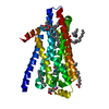
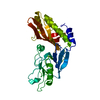
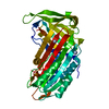
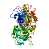
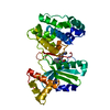
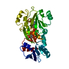
- PDB-5dhh: The crystal structure of nociceptin/orphanin FQ peptide receptor ... -
+ Open data

- Basic information
| Entry | Database: PDB / ID: 5dhh | ||||||
|---|---|---|---|---|---|---|---|
| Title | The crystal structure of nociceptin/orphanin FQ peptide receptor (NOP) in complex with SB-612111 (PSI Community Target) | ||||||
 Components Components | Soluble cytochrome b562,Nociceptin receptor | ||||||
 Keywords Keywords | SIGNALING PROTEIN / Nociceptin/orphanin FQ peptide receptor / NOP / ORL-1 / N/OFQ / opioid receptor / G protein-coupled receptor / GPCR / membrane protein / lipidic cubic phase / BRET / receptor-ligand conformational pair / Structural Genomics / PSI-Biology / GPCR Network / PSICNT-127 | ||||||
| Function / homology |  Function and homology information Function and homology informationnociceptin receptor activity / neuron-neuron synaptic transmission / neuropeptide binding / sensory perception / sensory perception of pain / Peptide ligand-binding receptors / synaptic membrane / calcium-mediated signaling / neuropeptide signaling pathway / electron transport chain ...nociceptin receptor activity / neuron-neuron synaptic transmission / neuropeptide binding / sensory perception / sensory perception of pain / Peptide ligand-binding receptors / synaptic membrane / calcium-mediated signaling / neuropeptide signaling pathway / electron transport chain / G protein-coupled receptor activity / adenylate cyclase-inhibiting G protein-coupled receptor signaling pathway / cytoplasmic vesicle / G alpha (i) signalling events / phospholipase C-activating G protein-coupled receptor signaling pathway / periplasmic space / electron transfer activity / neuron projection / iron ion binding / heme binding / plasma membrane Similarity search - Function | ||||||
| Biological species |   Homo sapiens (human) Homo sapiens (human) | ||||||
| Method |  X-RAY DIFFRACTION / SYNCHROTRON / MOLECULAR REPLACEMENT / Resolution: 3.004 Å X-RAY DIFFRACTION / SYNCHROTRON / MOLECULAR REPLACEMENT / Resolution: 3.004 Å | ||||||
 Authors Authors | Miller, R.L. / Thompson, A.A. / Trapella, C. / Guerrini, R. / Malfacini, D. / Patel, N. / Han, G.W. / Cherezov, V. / Calo, G. / Katritch, V. ...Miller, R.L. / Thompson, A.A. / Trapella, C. / Guerrini, R. / Malfacini, D. / Patel, N. / Han, G.W. / Cherezov, V. / Calo, G. / Katritch, V. / Stevens, R.C. / GPCR Network (GPCR) | ||||||
 Citation Citation | Journal: Structure / Year: 2015 Title: The Importance of Ligand-Receptor Conformational Pairs in Stabilization: Spotlight on the N/OFQ G Protein-Coupled Receptor. Authors: Miller, R.L. / Thompson, A.A. / Trapella, C. / Guerrini, R. / Malfacini, D. / Patel, N. / Han, G.W. / Cherezov, V. / Calo, G. / Katritch, V. / Stevens, R.C. | ||||||
| History |
|
- Structure visualization
| Structure viewer | Molecule: MolmilJmol/JSmol |
|---|
- Downloads & links
-Download
| PDBx/mmCIF format | 5dhh.cif.gz | 272.6 KB | Display | PDBx/mmCIF format |
|---|---|---|---|---|
| PDB format | pdb5dhh.ent.gz | 216.5 KB | Display | PDB format |
| PDBx/mmJSON format | 5dhh.json.gz | Tree view | PDBx/mmJSON format | |
| Others |  Other downloads Other downloads |
-Validation report
| Arichive directory | https://data.pdbj.org/pub/pdb/validation_reports/dh/5dhhftp://data.pdbj.org/pub/pdb/validation_reports/dh/5dhh | HTTPS FTP |
|---|
-Related structure data
| Related structure data |  5dhgC 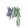 4ea3S S: Starting model for refinement C: citing same article ( |
|---|---|
| Similar structure data | |
| Other databases |


-Links
 PDBj
PDBj


- Assembly
| Deposited unit | 
| ||||||||
|---|---|---|---|---|---|---|---|---|---|
| 1 | 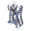
| ||||||||
| 2 | 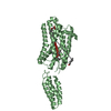
| ||||||||
| 3 | 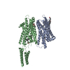
| ||||||||
| Unit cell |
|
-Components
| #1: Protein | Mass: 46696.371 Da / Num. of mol.: 2 / Mutation: M1007W, H1102I, R1106L Source method: isolated from a genetically manipulated source Source: (gene. exp.) Homo sapiens (human)Strain: O26:H11 / Gene: cybC, OPRL1, OOR, ORL1 / Plasmid: pFASTBAC / Cell line (production host): sf9 / Production host:   spodoptera frugiperda (fall armyworm) / Strain (production host): sf9 / References: UniProt: P0ABE7, UniProt: P41146 spodoptera frugiperda (fall armyworm) / Strain (production host): sf9 / References: UniProt: P0ABE7, UniProt: P41146#2: Chemical | ChemComp-OLA /   Mass: 282.461 Da / Num. of mol.: 7 / Source method: obtained synthetically / Formula: C18H34O2 Mass: 282.461 Da / Num. of mol.: 7 / Source method: obtained synthetically / Formula: C18H34O2#3: Chemical |   Mass: 418.399 Da / Num. of mol.: 2 / Source method: obtained synthetically / Formula: C24H29Cl2NO Mass: 418.399 Da / Num. of mol.: 2 / Source method: obtained synthetically / Formula: C24H29Cl2NO#4: Chemical | ChemComp-OLC / ( |   Mass: 356.540 Da / Num. of mol.: 1 / Source method: obtained synthetically / Formula: C21H40O4 Mass: 356.540 Da / Num. of mol.: 1 / Source method: obtained synthetically / Formula: C21H40O4Has protein modification | Y | |
|---|
-Experimental details
-Experiment
| Experiment | Method: X-RAY DIFFRACTION |
|---|
- Sample preparation
| Crystal | Density Matthews: 2.48 Å3/Da / Density % sol: 50.4 % |
|---|---|
| Crystal grow | Temperature: 293 K / Method: lipidic cubic phase / pH: 6.4 Details: 25-35% (V/V) PEG400, 130-200 MM POTASSIM SODIUM TARTRATE TETRAHYDRATE, 100 MM BIS-TRIS PROPANE, PH 6.4, LIPIDIC CUBIC PHASE, TEMPERATURE 293K |
-Data collection
| Diffraction | Mean temperature: 100 K |
|---|---|
| Diffraction source | Source: SYNCHROTRON / Site: APS  / Beamline: 23-ID-D / Wavelength: 1.033 Å / Beamline: 23-ID-D / Wavelength: 1.033 Å |
| Detector | Type: MARMOSAIC 300 mm CCD / Detector: CCD / Date: Dec 8, 2013 / Details: Another data collection date: 2014-02-20 |
| Radiation | Monochromator: mirrors / Protocol: SINGLE WAVELENGTH / Monochromatic (M) / Laue (L): M / Scattering type: x-ray |
| Radiation wavelength | Wavelength: 1.033 Å / Relative weight: 1 |
| Reflection | Resolution: 3→30 Å / Num. obs: 16552 / % possible obs: 93.2 % / Redundancy: 3.4 % / Rmerge(I) obs: 0.191 / Net I/σ(I): 7.27 |
| Reflection shell | Resolution: 3→3.1 Å / Redundancy: 3.3 % / Mean I/σ(I) obs: 1.19 / % possible all: 91.8 |
- Processing
| Software |
| ||||||||||||||||||||||||||||||||||||||||||||||||||||||||||||||||||||||||||||||||||||||||||||||||||||
|---|---|---|---|---|---|---|---|---|---|---|---|---|---|---|---|---|---|---|---|---|---|---|---|---|---|---|---|---|---|---|---|---|---|---|---|---|---|---|---|---|---|---|---|---|---|---|---|---|---|---|---|---|---|---|---|---|---|---|---|---|---|---|---|---|---|---|---|---|---|---|---|---|---|---|---|---|---|---|---|---|---|---|---|---|---|---|---|---|---|---|---|---|---|---|---|---|---|---|---|---|---|
| Refinement | Method to determine structure: MOLECULAR REPLACEMENT Starting model: 4EA3 Resolution: 3.004→29.845 Å / SU ML: 0.47 / Cross valid method: THROUGHOUT / σ(F): 1.34 / Phase error: 30.05 / Stereochemistry target values: ML
| ||||||||||||||||||||||||||||||||||||||||||||||||||||||||||||||||||||||||||||||||||||||||||||||||||||
| Solvent computation | Shrinkage radii: 0.9 Å / VDW probe radii: 1.11 Å / Solvent model: FLAT BULK SOLVENT MODEL | ||||||||||||||||||||||||||||||||||||||||||||||||||||||||||||||||||||||||||||||||||||||||||||||||||||
| Refinement step | Cycle: LAST / Resolution: 3.004→29.845 Å
| ||||||||||||||||||||||||||||||||||||||||||||||||||||||||||||||||||||||||||||||||||||||||||||||||||||
| Refine LS restraints |
| ||||||||||||||||||||||||||||||||||||||||||||||||||||||||||||||||||||||||||||||||||||||||||||||||||||
| LS refinement shell |
| ||||||||||||||||||||||||||||||||||||||||||||||||||||||||||||||||||||||||||||||||||||||||||||||||||||
| Refinement TLS params. | Method: refined / Refine-ID: X-RAY DIFFRACTION
| ||||||||||||||||||||||||||||||||||||||||||||||||||||||||||||||||||||||||||||||||||||||||||||||||||||
| Refinement TLS group |
|
