 Movie
Movie Controller

[English] 日本語
 Yorodumi
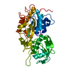
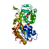
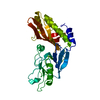
- PDB-4f1u: Subatomic resolution structure of a high affinity periplasmic pho... -
+ Open data

- Basic information
| Entry | Database: PDB / ID: 4f1u | |||||||||
|---|---|---|---|---|---|---|---|---|---|---|
| Title | Subatomic resolution structure of a high affinity periplasmic phosphate-binding protein (PfluDING) bound with phosphate at pH 4.5 | |||||||||
 Components Components | Putative alkaline phosphatase | |||||||||
 Keywords Keywords | PHOSPHATE-BINDING PROTEIN / venus flytrap / phosphate binding protein / pstS / DING | |||||||||
| Function / homology |  Function and homology information Function and homology informationphosphate ion transmembrane transport / phosphate ion binding / ATP-binding cassette (ABC) transporter complex Similarity search - Function | |||||||||
| Biological species |  Pseudomonas fluorescens (bacteria) Pseudomonas fluorescens (bacteria) | |||||||||
| Method |  X-RAY DIFFRACTION / SYNCHROTRON / MOLECULAR REPLACEMENT / Resolution: 0.98 Å X-RAY DIFFRACTION / SYNCHROTRON / MOLECULAR REPLACEMENT / Resolution: 0.98 Å | |||||||||
 Authors Authors | Liebschner, D. / Elias, M. / Tawfik, D.S. / Moniot, S. / Fournier, B. / Scott, K. / Jelsch, C. / Guillot, B. / Lecomte, C. / Chabriere, E. | |||||||||
 Citation Citation | Journal: Nature / Year: 2012 Title: The molecular basis of phosphate discrimination in arsenate-rich environments. Authors: Elias, M. / Wellner, A. / Goldin-Azulay, K. / Chabriere, E. / Vorholt, J.A. / Erb, T.J. / Tawfik, D.S. #1: Journal: J.Am.Chem.Soc. / Year: 2009 Title: Elucidation of the phosphate binding mode of DING proteins revealed by subangstrom X-ray crystallography. Authors: Liebschner, D. / Elias, M. / Moniot, S. / Fournier, B. / Scott, K. / Jelsch, C. / Guillot, B. / Lecomte, C. / Chabriere, E. | |||||||||
| History |
|
- Structure visualization
| Structure viewer | Molecule: MolmilJmol/JSmol |
|---|
- Downloads & links
-Download
| PDBx/mmCIF format | 4f1u.cif.gz | 369.2 KB | Display | PDBx/mmCIF format |
|---|---|---|---|---|
| PDB format | pdb4f1u.ent.gz | 303.3 KB | Display | PDB format |
| PDBx/mmJSON format | 4f1u.json.gz | Tree view | PDBx/mmJSON format | |
| Others |  Other downloads Other downloads |
-Validation report
| Arichive directory | https://data.pdbj.org/pub/pdb/validation_reports/f1/4f1uftp://data.pdbj.org/pub/pdb/validation_reports/f1/4f1u | HTTPS FTP |
|---|
-Related structure data
| Related structure data |  4f18C  4f19C 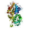 4f1vC  3g62 C: citing same article ( S: Starting model for refinement |
|---|---|
| Similar structure data |


-Links
 PDBj
PDBj- Assembly
| Deposited unit | 
| ||||||||
|---|---|---|---|---|---|---|---|---|---|
| 1 |
| ||||||||
| Unit cell |
|
-Components
| #1: Protein | Mass: 39119.289 Da / Num. of mol.: 1 Source method: isolated from a genetically manipulated source Source: (gene. exp.) Pseudomonas fluorescens (bacteria) / Strain: SBW25 / Gene: PFLU_2427 / Production host: | ||||||
|---|---|---|---|---|---|---|---|
| #2: Chemical | ChemComp-PI /   Mass: 95.979 Da / Num. of mol.: 1 / Source method: obtained synthetically / Formula: HO4P Mass: 95.979 Da / Num. of mol.: 1 / Source method: obtained synthetically / Formula: HO4P | ||||||
| #3: Chemical | ChemComp-EDO /   Mass: 62.068 Da / Num. of mol.: 4 / Source method: obtained synthetically / Formula: C2H6O2 Mass: 62.068 Da / Num. of mol.: 4 / Source method: obtained synthetically / Formula: C2H6O2#4: Chemical | ChemComp-SO4 / |   Mass: 96.063 Da / Num. of mol.: 1 / Source method: obtained synthetically / Formula: SO4 Mass: 96.063 Da / Num. of mol.: 1 / Source method: obtained synthetically / Formula: SO4#5: Water | ChemComp-HOH / |  Mass: 18.015 Da / Num. of mol.: 1234 / Source method: isolated from a natural source / Formula: H2O Mass: 18.015 Da / Num. of mol.: 1234 / Source method: isolated from a natural source / Formula: H2OHas protein modification | Y | |
-Experimental details
-Experiment
| Experiment | Method: X-RAY DIFFRACTION / Number of used crystals: 1 |
|---|
- Sample preparation
| Crystal | Density Matthews: 2.16 Å3/Da / Density % sol: 42.93 % |
|---|---|
| Crystal grow | Temperature: 293 K / Method: vapor diffusion, hanging drop / pH: 4.5 Details: 4% PEG 8000, 100 mM acetate buffer pH 4.5, 200 mM Li2SO4, VAPOR DIFFUSION, HANGING DROP, temperature 293.0K |
-Data collection
| Diffraction source | Source: SYNCHROTRON / Site: ESRF  / Beamline: ID29 / Wavelength: 0.953 Å / Beamline: ID29 / Wavelength: 0.953 Å |
|---|---|
| Detector | Type: ADSC QUANTUM 315 / Detector: CCD / Date: Dec 5, 2006 |
| Radiation | Protocol: SINGLE WAVELENGTH / Monochromatic (M) / Laue (L): M / Scattering type: x-ray |
| Radiation wavelength | Wavelength: 0.953 Å / Relative weight: 1 |
| Reflection | Resolution: 0.98→36.69 Å / Num. obs: 185558 / % possible obs: 98.1 % / Observed criterion σ(F): 2 / Observed criterion σ(I): 2 |
- Processing
| Software |
| ||||||||||||||||||||||||||||||||||||||||||||||||||||||||||||||||||||||||||||||||||||||||||||||||||||||||||||||||||||||||||||||||||||||||||||||||||||||||||||||||||||||||||
|---|---|---|---|---|---|---|---|---|---|---|---|---|---|---|---|---|---|---|---|---|---|---|---|---|---|---|---|---|---|---|---|---|---|---|---|---|---|---|---|---|---|---|---|---|---|---|---|---|---|---|---|---|---|---|---|---|---|---|---|---|---|---|---|---|---|---|---|---|---|---|---|---|---|---|---|---|---|---|---|---|---|---|---|---|---|---|---|---|---|---|---|---|---|---|---|---|---|---|---|---|---|---|---|---|---|---|---|---|---|---|---|---|---|---|---|---|---|---|---|---|---|---|---|---|---|---|---|---|---|---|---|---|---|---|---|---|---|---|---|---|---|---|---|---|---|---|---|---|---|---|---|---|---|---|---|---|---|---|---|---|---|---|---|---|---|---|---|---|---|---|---|
| Refinement | Method to determine structure: MOLECULAR REPLACEMENT Starting model: PDB ENTRY 3G62 3g62 Resolution: 0.98→36.69 Å / Cor.coef. Fo:Fc: 0.987 / Cor.coef. Fo:Fc free: 0.983 / SU B: 0.338 / SU ML: 0.009 / Cross valid method: THROUGHOUT / ESU R: 0.016 / ESU R Free: 0.015 / Stereochemistry target values: MAXIMUM LIKELIHOOD / Details: HYDROGENS HAVE BEEN USED IF PRESENT IN THE INPUT
| ||||||||||||||||||||||||||||||||||||||||||||||||||||||||||||||||||||||||||||||||||||||||||||||||||||||||||||||||||||||||||||||||||||||||||||||||||||||||||||||||||||||||||
| Solvent computation | Ion probe radii: 0.8 Å / Shrinkage radii: 0.8 Å / VDW probe radii: 1.2 Å / Solvent model: MASK | ||||||||||||||||||||||||||||||||||||||||||||||||||||||||||||||||||||||||||||||||||||||||||||||||||||||||||||||||||||||||||||||||||||||||||||||||||||||||||||||||||||||||||
| Displacement parameters | Biso mean: 8.948 Å2
| ||||||||||||||||||||||||||||||||||||||||||||||||||||||||||||||||||||||||||||||||||||||||||||||||||||||||||||||||||||||||||||||||||||||||||||||||||||||||||||||||||||||||||
| Refinement step | Cycle: LAST / Resolution: 0.98→36.69 Å
| ||||||||||||||||||||||||||||||||||||||||||||||||||||||||||||||||||||||||||||||||||||||||||||||||||||||||||||||||||||||||||||||||||||||||||||||||||||||||||||||||||||||||||
| Refine LS restraints |
| ||||||||||||||||||||||||||||||||||||||||||||||||||||||||||||||||||||||||||||||||||||||||||||||||||||||||||||||||||||||||||||||||||||||||||||||||||||||||||||||||||||||||||
| LS refinement shell | Resolution: 0.98→1.005 Å / Total num. of bins used: 20
|
