 Movie
Movie Controller

[English] 日本語
 Yorodumi
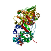
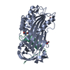
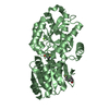
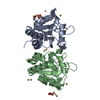
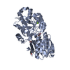
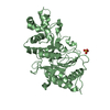
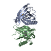
- PDB-2q9t: High-resolution structure of the DING protein from Pseudomonas fl... -
+ Open data

- Basic information
| Entry | Database: PDB / ID: 2q9t | ||||||
|---|---|---|---|---|---|---|---|
| Title | High-resolution structure of the DING protein from Pseudomonas fluorescens | ||||||
 Components Components | DING | ||||||
 Keywords Keywords | UNKNOWN FUNCTION / DING protein / phosphate-binding / Venus flytrap fold / PstS protein | ||||||
| Function / homology |  Function and homology information Function and homology informationphosphate ion transmembrane transport / phosphate ion binding / ATP-binding cassette (ABC) transporter complex / hydrolase activity Similarity search - Function | ||||||
| Biological species |  Pseudomonas fluorescens (bacteria) Pseudomonas fluorescens (bacteria) | ||||||
| Method |  X-RAY DIFFRACTION / SYNCHROTRON / MOLECULAR REPLACEMENT / Resolution: 1.43 Å X-RAY DIFFRACTION / SYNCHROTRON / MOLECULAR REPLACEMENT / Resolution: 1.43 Å | ||||||
 Authors Authors | Moniot, S. / Ahn, S. / Elias, M. / Kim, D. / Scott, K. / Chabriere, E. | ||||||
 Citation Citation | Journal: Febs Lett. / Year: 2007 Title: Structure-function relationships in a bacterial DING protein. Authors: Ahn, S. / Moniot, S. / Elias, M. / Chabriere, E. / Kim, D. / Scott, K. | ||||||
| History |
| ||||||
| Remark 999 | sequence The sequence of this protein in not available in the UNP database at the time of ...sequence The sequence of this protein in not available in the UNP database at the time of processing. The C-terminal six His residues are cloning artifacts. |
- Structure visualization
| Structure viewer | Molecule: MolmilJmol/JSmol |
|---|
- Downloads & links
-Download
| PDBx/mmCIF format | 2q9t.cif.gz | 184.2 KB | Display | PDBx/mmCIF format |
|---|---|---|---|---|
| PDB format | pdb2q9t.ent.gz | 144.2 KB | Display | PDB format |
| PDBx/mmJSON format | 2q9t.json.gz | Tree view | PDBx/mmJSON format | |
| Others |  Other downloads Other downloads |
-Validation report
| Arichive directory | https://data.pdbj.org/pub/pdb/validation_reports/q9/2q9tftp://data.pdbj.org/pub/pdb/validation_reports/q9/2q9t | HTTPS FTP |
|---|
-Related structure data
| Related structure data |  2cap S: Starting model for refinement |
|---|---|
| Similar structure data |


-Links
 PDBj
PDBj- Assembly
| Deposited unit | 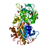
| ||||||||
|---|---|---|---|---|---|---|---|---|---|
| 1 |
| ||||||||
| Unit cell |
|
-Components
-Protein , 1 types, 1 molecules A
| #1: Protein | Mass: 38977.133 Da / Num. of mol.: 1 Source method: isolated from a genetically manipulated source Details: C-terminal hexahistidine tag / Source: (gene. exp.) Pseudomonas fluorescens (bacteria) / Strain: SBW 25 / Plasmid: pET22b(+) / Production host: |
|---|
-Non-polymers , 6 types, 777 molecules 


| #2: Chemical | ChemComp-PO4 /  Mass: 94.971 Da / Num. of mol.: 1 / Source method: obtained synthetically / Formula: PO4 Mass: 94.971 Da / Num. of mol.: 1 / Source method: obtained synthetically / Formula: PO4 | ||||||||
|---|---|---|---|---|---|---|---|---|---|
| #3: Chemical |  Mass: 59.044 Da / Num. of mol.: 2 / Source method: obtained synthetically / Formula: C2H3O2 Mass: 59.044 Da / Num. of mol.: 2 / Source method: obtained synthetically / Formula: C2H3O2#4: Chemical |  Mass: 96.063 Da / Num. of mol.: 2 / Source method: obtained synthetically / Formula: SO4 Mass: 96.063 Da / Num. of mol.: 2 / Source method: obtained synthetically / Formula: SO4#5: Chemical | ChemComp-EDO /  Mass: 62.068 Da / Num. of mol.: 5 / Source method: obtained synthetically / Formula: C2H6O2 Mass: 62.068 Da / Num. of mol.: 5 / Source method: obtained synthetically / Formula: C2H6O2#6: Chemical | ChemComp-GOL / |  Mass: 92.094 Da / Num. of mol.: 1 / Source method: obtained synthetically / Formula: C3H8O3 Mass: 92.094 Da / Num. of mol.: 1 / Source method: obtained synthetically / Formula: C3H8O3#7: Water | ChemComp-HOH / | Mass: 18.015 Da / Num. of mol.: 766 / Source method: isolated from a natural source / Formula: H2O |
-Details
| Has protein modification | Y |
|---|
-Experimental details
-Experiment
| Experiment | Method: X-RAY DIFFRACTION / Number of used crystals: 1 |
|---|
- Sample preparation
| Crystal | Density Matthews: 2.12 Å3/Da / Density % sol: 42.1 % |
|---|---|
| Crystal grow | Temperature: 293 K / Method: vapor diffusion, hanging drop / pH: 4.5 Details: 24% PEG 8000, 100 mM acetate buffer pH 4.5, 200 mM Li2SO4, VAPOR DIFFUSION, HANGING DROP, temperature 293K |
-Data collection
| Diffraction | Mean temperature: 100 K |
|---|---|
| Diffraction source | Source: SYNCHROTRON / Site: ESRF  / Beamline: BM14 / Wavelength: 0.9535 Å / Beamline: BM14 / Wavelength: 0.9535 Å |
| Detector | Type: MARMOSAIC 225 mm CCD / Detector: CCD / Date: Jul 9, 2006 |
| Radiation | Protocol: SINGLE WAVELENGTH / Monochromatic (M) / Laue (L): M / Scattering type: x-ray |
| Radiation wavelength | Wavelength: 0.9535 Å / Relative weight: 1 |
| Reflection | Resolution: 1.43→61.9 Å / Num. all: 60045 / Num. obs: 59063 / % possible obs: 98.1 % / Redundancy: 4 % / Biso Wilson estimate: 8.9 Å2 / Rsym value: 0.026 / Net I/σ(I): 38.9 |
| Reflection shell | Resolution: 1.43→1.5 Å / Redundancy: 2.9 % / Mean I/σ(I) obs: 10.52 / Num. unique all: 7166 / Rsym value: 0.102 / % possible all: 89.2 |
- Processing
| Software |
| |||||||||||||||||||||||||||||||||||||||||||||||||||||||||||||||||||||||||||||||||||||||||||||||||||||||||
|---|---|---|---|---|---|---|---|---|---|---|---|---|---|---|---|---|---|---|---|---|---|---|---|---|---|---|---|---|---|---|---|---|---|---|---|---|---|---|---|---|---|---|---|---|---|---|---|---|---|---|---|---|---|---|---|---|---|---|---|---|---|---|---|---|---|---|---|---|---|---|---|---|---|---|---|---|---|---|---|---|---|---|---|---|---|---|---|---|---|---|---|---|---|---|---|---|---|---|---|---|---|---|---|---|---|---|
| Refinement | Method to determine structure: MOLECULAR REPLACEMENT Starting model: Human Phosphate-binding protein (HPBP) PDB code 2cap 2cap Resolution: 1.43→61.9 Å / Cor.coef. Fo:Fc: 0.972 / Cor.coef. Fo:Fc free: 0.956 / SU B: 1.818 / SU ML: 0.034 / Isotropic thermal model: anisotropic / Cross valid method: THROUGHOUT / σ(F): 0 / ESU R: 0.078 / ESU R Free: 0.062 / Stereochemistry target values: MAXIMUM LIKELIHOOD / Details: HYDROGENS HAVE BEEN ADDED IN THE RIDING POSITIONS
| |||||||||||||||||||||||||||||||||||||||||||||||||||||||||||||||||||||||||||||||||||||||||||||||||||||||||
| Solvent computation | Ion probe radii: 0.8 Å / Shrinkage radii: 0.8 Å / VDW probe radii: 1.2 Å / Solvent model: MASK | |||||||||||||||||||||||||||||||||||||||||||||||||||||||||||||||||||||||||||||||||||||||||||||||||||||||||
| Displacement parameters | Biso mean: 10.155 Å2
| |||||||||||||||||||||||||||||||||||||||||||||||||||||||||||||||||||||||||||||||||||||||||||||||||||||||||
| Refinement step | Cycle: LAST / Resolution: 1.43→61.9 Å
| |||||||||||||||||||||||||||||||||||||||||||||||||||||||||||||||||||||||||||||||||||||||||||||||||||||||||
| Refine LS restraints |
| |||||||||||||||||||||||||||||||||||||||||||||||||||||||||||||||||||||||||||||||||||||||||||||||||||||||||
| LS refinement shell | Resolution: 1.43→1.467 Å / Total num. of bins used: 20
|
