 Movie
Movie Controller

[English] 日本語
 Yorodumi
- PDB-5nqf: Crystal structure of Plasmodium falciparum AMA1 in complex with a... -
+ Open data

- Basic information
| Entry | Database: PDB / ID: 5nqf | ||||||
|---|---|---|---|---|---|---|---|
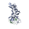
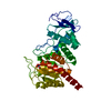
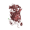
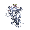
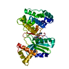
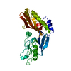
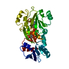
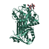
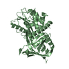
| Title | Crystal structure of Plasmodium falciparum AMA1 in complex with a 39 aa PvRON2 peptide | ||||||
 Components Components |
| ||||||
 Keywords Keywords | CELL INVASION / AMA1 / RON2 / MALARIA | ||||||
| Function / homology |  Function and homology information Function and homology information | ||||||
| Biological species |  | ||||||
| Method |  X-RAY DIFFRACTION / SYNCHROTRON / MOLECULAR REPLACEMENT / Resolution: 1.9 Å X-RAY DIFFRACTION / SYNCHROTRON / MOLECULAR REPLACEMENT / Resolution: 1.9 Å | ||||||
 Authors Authors | Vulliez-le Normand, B. / Saul, F.A. / Faber, B.W. / Bentley, G.A. | ||||||
 Citation Citation | Journal: PLoS ONE / Year: 2017 Title: Cross-reactivity between apical membrane antgen 1 and rhoptry neck protein 2 in P. vivax and P. falciparum: A structural and binding study. Authors: Vulliez-Le Normand, B. / Saul, F.A. / Hoos, S. / Faber, B.W. / Bentley, G.A. | ||||||
| History |
|
- Structure visualization
| Structure viewer | Molecule: MolmilJmol/JSmol |
|---|
- Downloads & links
-Download
| PDBx/mmCIF format | 5nqf.cif.gz | 91.6 KB | Display | PDBx/mmCIF format |
|---|---|---|---|---|
| PDB format | pdb5nqf.ent.gz | 65.1 KB | Display | PDB format |
| PDBx/mmJSON format | 5nqf.json.gz | Tree view | PDBx/mmJSON format | |
| Others |  Other downloads Other downloads |
-Validation report
| Arichive directory | https://data.pdbj.org/pub/pdb/validation_reports/nq/5nqfftp://data.pdbj.org/pub/pdb/validation_reports/nq/5nqf | HTTPS FTP |
|---|
-Related structure data

-Links
 PDBj
PDBj- Assembly
| Deposited unit | 
| ||||||||
|---|---|---|---|---|---|---|---|---|---|
| 1 |
| ||||||||
| Unit cell |
|
-Components
| #1: Protein | Mass: 42690.598 Da / Num. of mol.: 1 / Fragment: UNP RESIDUES 97-442 / Mutation: YES Source method: isolated from a genetically manipulated source Details: RESIDUES I97-P442 OF PFAMA1-FVO. N-TERMINAL STRETCH EF IS A CLONING ARTEFACT. C-TERMINAL STRETCH GLEQKLISEEDLNSAVDHHHHHH IS AN EXPRESSION TAG ( C-MYC + HEXA-HIS ). N162K, T288V, S373D, ...Details: RESIDUES I97-P442 OF PFAMA1-FVO. N-TERMINAL STRETCH EF IS A CLONING ARTEFACT. C-TERMINAL STRETCH GLEQKLISEEDLNSAVDHHHHHH IS AN EXPRESSION TAG ( C-MYC + HEXA-HIS ). N162K, T288V, S373D, N422D, S423K ARE ENGINEERED MUTATIONS. Source: (gene. exp.) Gene: PFFVO_05649 / Production host:  Komagataella pastoris (fungus) / References: UniProt: A0A024UZE1 Komagataella pastoris (fungus) / References: UniProt: A0A024UZE1 |
|---|---|
| #2: Protein/peptide | Mass: 4106.740 Da / Num. of mol.: 1 / Fragment: UNP Residues 2034-2072 / Source method: obtained synthetically / Source: (synth.) |
| #3: Water | ChemComp-HOH /  Mass: 18.015 Da / Num. of mol.: 307 / Source method: isolated from a natural source / Formula: H2O Mass: 18.015 Da / Num. of mol.: 307 / Source method: isolated from a natural source / Formula: H2O |
| Has protein modification | Y |
-Experimental details
-Experiment
| Experiment | Method: X-RAY DIFFRACTION / Number of used crystals: 1 |
|---|
- Sample preparation
| Crystal | Density Matthews: 2.1 Å3/Da / Density % sol: 41.3 % |
|---|---|
| Crystal grow | Temperature: 290 K / Method: vapor diffusion, sitting drop / pH: 8.2 Details: PEG 3350, TRIS 8.2, SODIUM ACETATE, ISOPROPANOL, PH 8.2, VAPOR DIFFUSION, SITTING DROP, TEMPERATURE 290K PH range: 8.2 |
-Data collection
| Diffraction | Mean temperature: 110 K |
|---|---|
| Diffraction source | Source: SYNCHROTRON / Site: SOLEIL  / Beamline: PROXIMA 1 / Wavelength: 0.91165 Å / Beamline: PROXIMA 1 / Wavelength: 0.91165 Å |
| Detector | Type: DECTRIS PILATUS 6M / Detector: PIXEL / Date: Apr 4, 2013 |
| Radiation | Monochromator: SI(111) / Protocol: SINGLE WAVELENGTH / Monochromatic (M) / Laue (L): M / Scattering type: x-ray |
| Radiation wavelength | Wavelength: 0.91165 Å / Relative weight: 1 |
| Reflection | Resolution: 1.9→46.92 Å / Num. obs: 29741 / % possible obs: 96.2 % / Redundancy: 3 % / Biso Wilson estimate: 23.33 Å2 / CC1/2: 0.996 / Rmerge(I) obs: 0.083 / Net I/σ(I): 9.2 |
| Reflection shell | Resolution: 1.9→2 Å / Redundancy: 1.8 % / Rmerge(I) obs: 0.569 / Mean I/σ(I) obs: 1.4 / CC1/2: 0.822 / % possible all: 78.3 |
- Processing
| Software |
| ||||||||||||||||||||||||||||||||||||||||||||||||||||||||||||||||||||||||||||||||||||||||||||||||||||||||||||||||||
|---|---|---|---|---|---|---|---|---|---|---|---|---|---|---|---|---|---|---|---|---|---|---|---|---|---|---|---|---|---|---|---|---|---|---|---|---|---|---|---|---|---|---|---|---|---|---|---|---|---|---|---|---|---|---|---|---|---|---|---|---|---|---|---|---|---|---|---|---|---|---|---|---|---|---|---|---|---|---|---|---|---|---|---|---|---|---|---|---|---|---|---|---|---|---|---|---|---|---|---|---|---|---|---|---|---|---|---|---|---|---|---|---|---|---|---|
| Refinement | Method to determine structure: MOLECULAR REPLACEMENT Starting model: AMA1-FVO Resolution: 1.9→35.78 Å / Cor.coef. Fo:Fc: 0.941 / Cor.coef. Fo:Fc free: 0.922 / SU R Cruickshank DPI: 0.132 / Cross valid method: THROUGHOUT / σ(F): 0 / SU R Blow DPI: 0.142 / SU Rfree Blow DPI: 0.128 / SU Rfree Cruickshank DPI: 0.124
| ||||||||||||||||||||||||||||||||||||||||||||||||||||||||||||||||||||||||||||||||||||||||||||||||||||||||||||||||||
| Displacement parameters | Biso mean: 28.57 Å2
| ||||||||||||||||||||||||||||||||||||||||||||||||||||||||||||||||||||||||||||||||||||||||||||||||||||||||||||||||||
| Refine analyze | Luzzati coordinate error obs: 0.214 Å | ||||||||||||||||||||||||||||||||||||||||||||||||||||||||||||||||||||||||||||||||||||||||||||||||||||||||||||||||||
| Refinement step | Cycle: LAST / Resolution: 1.9→35.78 Å
| ||||||||||||||||||||||||||||||||||||||||||||||||||||||||||||||||||||||||||||||||||||||||||||||||||||||||||||||||||
| Refine LS restraints |
| ||||||||||||||||||||||||||||||||||||||||||||||||||||||||||||||||||||||||||||||||||||||||||||||||||||||||||||||||||
| LS refinement shell | Resolution: 1.9→1.97 Å / Total num. of bins used: 15
|
