 Movie
Movie Controller


+ Open data

- Basic information
| Entry | Database: PDB / ID: 3srj | ||||||
|---|---|---|---|---|---|---|---|
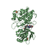
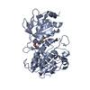
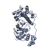
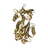
| Title | PfAMA1 in complex with invasion-inhibitory peptide R1 | ||||||
 Components Components |
| ||||||
 Keywords Keywords | CELL INVASION/inhibitor / AMA1 / Plasmodium falciparum / inhibitory peptide / malaria / CELL INVASION / CELL INVASION-inhibitor complex | ||||||
| Function / homology |  Function and homology information Function and homology informationapical complex / microneme / host cell surface binding / symbiont entry into host / membrane Similarity search - Function | ||||||
| Biological species |  | ||||||
| Method |  X-RAY DIFFRACTION / SYNCHROTRON / MOLECULAR REPLACEMENT / Resolution: 2.15 Å X-RAY DIFFRACTION / SYNCHROTRON / MOLECULAR REPLACEMENT / Resolution: 2.15 Å | ||||||
 Authors Authors | Vulliez-Le Normand, B. / Saul, F.A. / Bentley, G.A. | ||||||
 Citation Citation | Journal: Plos Pathog. / Year: 2012 Title: Structural and functional insights into the malaria parasite moving junction complex. Authors: Vulliez-Le Normand, B. / Tonkin, M.L. / Lamarque, M.H. / Langer, S. / Hoos, S. / Roques, M. / Saul, F.A. / Faber, B.W. / Bentley, G.A. / Boulanger, M.J. / Lebrun, M. | ||||||
| History |
|
- Structure visualization
| Structure viewer | Molecule: MolmilJmol/JSmol |
|---|
- Downloads & links
-Download
| PDBx/mmCIF format | 3srj.cif.gz | 155.7 KB | Display | PDBx/mmCIF format |
|---|---|---|---|---|
| PDB format | pdb3srj.ent.gz | 119.6 KB | Display | PDB format |
| PDBx/mmJSON format | 3srj.json.gz | Tree view | PDBx/mmJSON format | |
| Others |  Other downloads Other downloads |
-Validation report
| Arichive directory | https://data.pdbj.org/pub/pdb/validation_reports/sr/3srjftp://data.pdbj.org/pub/pdb/validation_reports/sr/3srj | HTTPS FTP |
|---|
-Related structure data
| Related structure data | 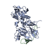 3sriC 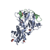 3zwzC 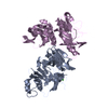 1z40S S: Starting model for refinement C: citing same article ( |
|---|---|
| Similar structure data |


-Links
 PDBj
PDBj- Assembly
| Deposited unit | 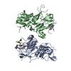
| ||||||||
|---|---|---|---|---|---|---|---|---|---|
| 1 | 
| ||||||||
| 2 | 
| ||||||||
| Unit cell |
|
-Components
| #1: Protein | Mass: 43665.910 Da / Num. of mol.: 2 / Fragment: AMA1 / Mutation: N162K, T288V, S373D, N422D, S423K Source method: isolated from a genetically manipulated source Source: (gene. exp.) Strain: 3D7 / Gene: AMA1, PF11_0344 / Production host:  Pichia pastoris (fungus) / References: UniProt: Q7KQK5 Pichia pastoris (fungus) / References: UniProt: Q7KQK5#2: Protein/peptide | Mass: 2371.883 Da / Num. of mol.: 4 / Source method: obtained synthetically #3: Water | ChemComp-HOH / |  Mass: 18.015 Da / Num. of mol.: 450 / Source method: isolated from a natural source / Formula: H2O Mass: 18.015 Da / Num. of mol.: 450 / Source method: isolated from a natural source / Formula: H2OHas protein modification | Y | |
|---|
-Experimental details
-Experiment
| Experiment | Method: X-RAY DIFFRACTION / Number of used crystals: 1 |
|---|
- Sample preparation
| Crystal | Density Matthews: 2.08 Å3/Da / Density % sol: 40.86 % |
|---|---|
| Crystal grow | Temperature: 290 K / Method: vapor diffusion, hanging drop / pH: 8.5 Details: 15% PEG 4000, 0.1M Tris/HCL, 0.1M sodium acetate, 10% isopropanol, pH 8.5, VAPOR DIFFUSION, HANGING DROP, temperature 290K |
-Data collection
| Diffraction | Mean temperature: 110 K |
|---|---|
| Diffraction source | Source: SYNCHROTRON / Site: SOLEIL  / Beamline: PROXIMA 1 / Wavelength: 0.97911 Å / Beamline: PROXIMA 1 / Wavelength: 0.97911 Å |
| Detector | Type: ADSC QUANTUM 315r / Detector: CCD / Date: Sep 17, 2010 |
| Radiation | Monochromator: Si 111 CHANNEL / Protocol: SINGLE WAVELENGTH / Monochromatic (M) / Laue (L): M / Scattering type: x-ray |
| Radiation wavelength | Wavelength: 0.97911 Å / Relative weight: 1 |
| Reflection | Resolution: 2.15→40.28 Å / Num. all: 42798 / Num. obs: 42798 / % possible obs: 95.3 % / Observed criterion σ(F): 0 / Observed criterion σ(I): -3 / Redundancy: 3.7 % / Biso Wilson estimate: 35.74 Å2 / Rmerge(I) obs: 0.075 / Net I/σ(I): 13.3 |
| Reflection shell | Resolution: 2.15→2.27 Å / Redundancy: 2.5 % / Rmerge(I) obs: 0.485 / Mean I/σ(I) obs: 2.2 / Num. unique all: 4756 / % possible all: 75.7 |
- Processing
| Software |
| ||||||||||||||||||||||||||||||||||||||||||||||||||||||||||||
|---|---|---|---|---|---|---|---|---|---|---|---|---|---|---|---|---|---|---|---|---|---|---|---|---|---|---|---|---|---|---|---|---|---|---|---|---|---|---|---|---|---|---|---|---|---|---|---|---|---|---|---|---|---|---|---|---|---|---|---|---|---|
| Refinement | Method to determine structure: MOLECULAR REPLACEMENT Starting model: PDB entry 1Z40 Resolution: 2.15→37.06 Å / Cor.coef. Fo:Fc: 0.9461 / Cor.coef. Fo:Fc free: 0.9173 / SU R Cruickshank DPI: 0.198 / Cross valid method: THROUGHOUT / σ(F): 0 / Stereochemistry target values: Engh & Huber
| ||||||||||||||||||||||||||||||||||||||||||||||||||||||||||||
| Displacement parameters | Biso mean: 41.15 Å2
| ||||||||||||||||||||||||||||||||||||||||||||||||||||||||||||
| Refine analyze | Luzzati coordinate error obs: 0.228 Å | ||||||||||||||||||||||||||||||||||||||||||||||||||||||||||||
| Refinement step | Cycle: LAST / Resolution: 2.15→37.06 Å
| ||||||||||||||||||||||||||||||||||||||||||||||||||||||||||||
| Refine LS restraints |
| ||||||||||||||||||||||||||||||||||||||||||||||||||||||||||||
| LS refinement shell | Resolution: 2.15→2.21 Å / Total num. of bins used: 20
|
