 Movie
Movie Controller

[English] 日本語
 Yorodumi
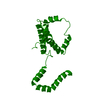
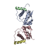
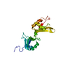
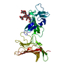
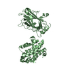
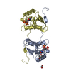
- PDB-3fkz: X-ray structure of the non covalent swapped form of the S16G/T17N... -
+ Open data

- Basic information
| Entry | Database: PDB / ID: 3fkz | ||||||
|---|---|---|---|---|---|---|---|
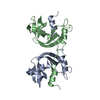
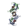
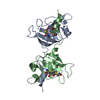
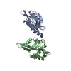
| Title | X-ray structure of the non covalent swapped form of the S16G/T17N/A19P/A20S/K31C/S32C mutant of bovine pancreatic ribonuclease | ||||||
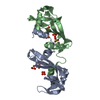
 Components Components | Ribonuclease pancreatic | ||||||
 Keywords Keywords | HYDROLASE / 3D-domain swapping / bovine seminal ribonuclease / non-covalent dimer / antitumor activity / quaternary structure flexibility / protein mutations and evolution / Endonuclease / Glycation / Glycoprotein / Nuclease / Secreted | ||||||
| Function / homology |  Function and homology information Function and homology informationpancreatic ribonuclease / ribonuclease A activity / RNA nuclease activity / nucleic acid binding / defense response to Gram-positive bacterium / hydrolase activity / extracellular region Similarity search - Function | ||||||
| Biological species |  | ||||||
| Method |  X-RAY DIFFRACTION / MOLECULAR REPLACEMENT / Resolution: 1.99 Å X-RAY DIFFRACTION / MOLECULAR REPLACEMENT / Resolution: 1.99 Å | ||||||
 Authors Authors | Merlino, A. / Russo Krauss, I. / Perillo, M. / Mattia, C.A. / Ercole, C. / Picone, D. / Vergara, A. / Sica, F. | ||||||
 Citation Citation | Journal: Biopolymers / Year: 2009 Title: Toward an antitumor form of bovine pancreatic ribonuclease: The crystal structure of three noncovalent dimeric mutants Authors: Merlino, A. / Russo Krauss, I. / Perillo, M. / Mattia, C.A. / Ercole, C. / Picone, D. / Vergara, A. / Sica, F. #1: Journal: J.Biol.Chem. / Year: 2004Title: Structure and stability of the non-covalent swapped dimer of bovine seminal ribonuclease: an enzyme tailored to evade ribonuclease protein inhibitor Authors: Sica, F. / Di Fiore, A. / Merlino, A. / Mazzarella, L. #2: Journal: J.Mol.Biol. / Year: 2008Title: The buried diversity of bovine seminal ribonuclease: shape and cytotoxicity of the swapped non-covalent form of the enzyme Authors: Merlino, A. / Ercole, C. / Picone, D. / Pizzo, E. / Mazzarella, L. / Sica, F. #3: Journal: Protein Sci. / Year: 1995 Title: Hints on the evolutionary design of a dimeric RNase with special bioactions Authors: Di Donato, A. / Cafaro, V. / Romeo, I. / D'Alessio, G. | ||||||
| History |
|
- Structure visualization
| Structure viewer | Molecule: MolmilJmol/JSmol |
|---|
- Downloads & links
-Download
| PDBx/mmCIF format | 3fkz.cif.gz | 62.7 KB | Display | PDBx/mmCIF format |
|---|---|---|---|---|
| PDB format | pdb3fkz.ent.gz | 46.7 KB | Display | PDB format |
| PDBx/mmJSON format | 3fkz.json.gz | Tree view | PDBx/mmJSON format | |
| Others |  Other downloads Other downloads |
-Validation report
| Arichive directory | https://data.pdbj.org/pub/pdb/validation_reports/fk/3fkzftp://data.pdbj.org/pub/pdb/validation_reports/fk/3fkz | HTTPS FTP |
|---|
-Related structure data
| Related structure data | 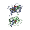 3fl0C 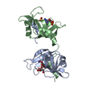 3fl1C 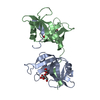 3fl3C  1kf3S C: citing same article ( S: Starting model for refinement |
|---|---|
| Similar structure data |


-Links
 PDBj
PDBj

- Assembly
| Deposited unit | 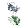
| ||||||||
|---|---|---|---|---|---|---|---|---|---|
| 1 |
| ||||||||
| Unit cell |
|
-Components
| #1: Protein | Mass: 13837.467 Da / Num. of mol.: 2 / Mutation: S16G, T17N, A19P, A20S, K31C, S32C Source method: isolated from a genetically manipulated source Source: (gene. exp.)  #2: Water | ChemComp-HOH / |  Mass: 18.015 Da / Num. of mol.: 122 / Source method: isolated from a natural source / Formula: H2O Mass: 18.015 Da / Num. of mol.: 122 / Source method: isolated from a natural source / Formula: H2O |
|---|
-Experimental details
-Experiment
| Experiment | Method: X-RAY DIFFRACTION / Number of used crystals: 1 |
|---|
- Sample preparation
| Crystal | Density Matthews: 2.32 Å3/Da / Density % sol: 46.9 % |
|---|---|
| Crystal grow | Temperature: 293 K / Method: vapor diffusion, hanging drop / pH: 8.5 Details: 30% PEG 4000, 0.2M lithium sulfate, 0.1M Tris/HCl, pH 8.5, VAPOR DIFFUSION, HANGING DROP, temperature 293K |
-Data collection
| Diffraction | Mean temperature: 100 K |
|---|---|
| Diffraction source | Source: ROTATING ANODE / Type: RIGAKU / Wavelength: 1.5418 Å |
| Detector | Type: RIGAKU SATURN 944 / Detector: CCD / Date: Apr 9, 2008 |
| Radiation | Monochromator: GRAPHITE / Protocol: SINGLE WAVELENGTH / Monochromatic (M) / Laue (L): M / Scattering type: x-ray |
| Radiation wavelength | Wavelength: 1.5418 Å / Relative weight: 1 |
| Reflection | Resolution: 1.99→50 Å / Num. all: 15901 / Num. obs: 15901 / % possible obs: 90.7 % / Observed criterion σ(F): 0 / Observed criterion σ(I): 0 / Redundancy: 6 % / Rmerge(I) obs: 0.08 / Net I/σ(I): 21 |
| Reflection shell | Resolution: 1.99→2.06 Å / Rmerge(I) obs: 0.285 / Mean I/σ(I) obs: 5 / % possible all: 56.5 |
- Processing
| Software |
| ||||||||||||||||||||
|---|---|---|---|---|---|---|---|---|---|---|---|---|---|---|---|---|---|---|---|---|---|
| Refinement | Method to determine structure: MOLECULAR REPLACEMENT Starting model: PDB ENTRY 1KF3 Resolution: 1.99→28.73 Å / Cross valid method: THROUGHOUT / σ(F): 2 / Stereochemistry target values: Engh & Huber
| ||||||||||||||||||||
| Refinement step | Cycle: LAST / Resolution: 1.99→28.73 Å
| ||||||||||||||||||||
| Refine LS restraints |
|
