- PDB-3etv: Crystal structure of a Tip20p-Dsl1p fusion protein -
+
Open data
ID or keywords:
Loading...
-
Basic information
Entry
Database: PDB / ID: 3etv
Title
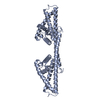
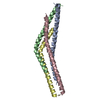
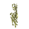
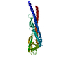
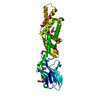
Crystal structure of a Tip20p-Dsl1p fusion protein
Components
Protein transport protein TIP20, Protein transport protein DSL1 chimera
Keywords
TRANSPORT PROTEIN / Tip20p-Dsl1p complex / Endoplasmic reticulum / ER-Golgi transport / Membrane / Phosphoprotein / Protein transport / Transport
Function / homology
Function and homology information
ER-dependent peroxisome organization / Dsl1/NZR complex / RZZ complex / regulation of ER to Golgi vesicle-mediated transport / COPI-dependent Golgi-to-ER retrograde traffic / retrograde vesicle-mediated transport, Golgi to endoplasmic reticulum / mitotic spindle assembly checkpoint signaling / endoplasmic reticulum to Golgi vesicle-mediated transport / autophagy / protein transport ...ER-dependent peroxisome organization / Dsl1/NZR complex / RZZ complex / regulation of ER to Golgi vesicle-mediated transport / COPI-dependent Golgi-to-ER retrograde traffic / retrograde vesicle-mediated transport, Golgi to endoplasmic reticulum / mitotic spindle assembly checkpoint signaling / endoplasmic reticulum to Golgi vesicle-mediated transport / autophagy / protein transport / endoplasmic reticulum membrane / endoplasmic reticulum / nucleus / cytosol Similarity search - Function
Helix Hairpins - #3290 / Retrograde transport protein Dsl1, N-terminal domain / RINT-1/Tip20 / Protein transport protein Tip20, domain E / Protein transport protein Tip20, domain A / Protein transport protein Tip20, domain B / Protein transport protein Tip20, domain C / RINT-1/TIP-1 family / RINT1/TIP20 domain profile. / Retrograde transport protein Dsl1 N-terminal domain ...Helix Hairpins - #3290 / Retrograde transport protein Dsl1, N-terminal domain / RINT-1/Tip20 / Protein transport protein Tip20, domain E / Protein transport protein Tip20, domain A / Protein transport protein Tip20, domain B / Protein transport protein Tip20, domain C / RINT-1/TIP-1 family / RINT1/TIP20 domain profile. / Retrograde transport protein Dsl1 N-terminal domain / Retrograde transport protein Dsl1, C-terminal domain / Dsl1, N-terminal domain superfamily / Retrograde transport protein Dsl1 N terminal / Retrograde transport protein Dsl1 C terminal / EXOC6/PINT-1/Sec15/Tip20, C-terminal, domain 2 / Zw10/DSL1, C-terminal / Methane Monooxygenase Hydroxylase; Chain G, domain 1 / Helix Hairpins / Up-down Bundle / Orthogonal Bundle / Mainly Alpha Similarity search - Domain/homology
Mass: 18.015 Da / Num. of mol.: 221 / Source method: isolated from a natural source / Formula: H2O
Sequence details
N-TERMINAL HELIX OF TIP20P IS FUSED TO DSL1P VIA AN EIGHT-RESIDUE GGGSGGGS LINKER
-
Experimental details
-
Experiment
Experiment
Method: X-RAY DIFFRACTION / Number of used crystals: 1
-
Sample preparation
Crystal
Density Matthews: 2.39 Å3/Da / Density % sol: 48.48 %
Crystal grow
Temperature: 298 K / Method: vapor diffusion / pH: 5 Details: 5:5:1 ratio of protein, well buffer (0.1 M sodium acetate, pH 5.0, 0.2 M ammonium acetate, 20% (w/v) PEG 4000), and additive (1.0 M lithium chloride) , VAPOR DIFFUSION, temperature 298.0K
Monochromator: Double silicon(111) crystal monochromator with cryogenically-cooled first crystal and sagittally-bent second crystal horizontally-focusing at 3.3:1 demagnification Protocol: SINGLE WAVELENGTH / Monochromatic (M) / Laue (L): M / Scattering type: x-ray
Radiation wavelength
Wavelength: 1.04 Å / Relative weight: 1
Reflection
Resolution: 1.94→100 Å / Num. obs: 25475 / % possible obs: 90.7 % / Redundancy: 3.1 % / Rsym value: 0.057 / Net I/σ(I): 18
Reflection shell
Resolution: 1.94→2.02 Å / Redundancy: 2.9 % / Mean I/σ(I) obs: 2 / Num. unique all: 2691 / Rsym value: 0.467 / % possible all: 97
Resolution: 1.94→40 Å / Cor.coef. Fo:Fc: 0.937 / Cor.coef. Fo:Fc free: 0.907 / SU B: 4.97 / SU ML: 0.143 / Cross valid method: THROUGHOUT / ESU R: 0.212 / ESU R Free: 0.192 / Stereochemistry target values: MAXIMUM LIKELIHOOD / Details: HYDROGENS HAVE BEEN ADDED IN THE RIDING POSITIONS
Rfactor
Num. reflection
% reflection
Selection details
Rfree
0.27722
1281
5 %
RANDOM
Rwork
0.22453
-
-
-
obs
0.22737
24166
90.35 %
-
Solvent computation
Ion probe radii: 0.8 Å / Shrinkage radii: 0.8 Å / VDW probe radii: 1.2 Å / Solvent model: MASK
Displacement parameters
Biso mean: 28.559 Å2
Baniso -1
Baniso -2
Baniso -3
1-
0.73 Å2
0 Å2
0.11 Å2
2-
-
-1.35 Å2
0 Å2
3-
-
-
0.63 Å2
Refinement step
Cycle: LAST / Resolution: 1.94→40 Å
Protein
Nucleic acid
Ligand
Solvent
Total
Num. atoms
2566
0
0
221
2787
Refine LS restraints
Refine-ID
Type
Dev ideal
Dev ideal target
Number
X-RAY DIFFRACTION
r_bond_refined_d
0.009
0.022
2605
X-RAY DIFFRACTION
r_bond_other_d
X-RAY DIFFRACTION
r_angle_refined_deg
1.091
1.958
3512
X-RAY DIFFRACTION
r_angle_other_deg
X-RAY DIFFRACTION
r_dihedral_angle_1_deg
5.44
5
312
X-RAY DIFFRACTION
r_dihedral_angle_2_deg
36.886
25.68
125
X-RAY DIFFRACTION
r_dihedral_angle_3_deg
13.308
15
508
X-RAY DIFFRACTION
r_dihedral_angle_4_deg
20.888
15
10
X-RAY DIFFRACTION
r_chiral_restr
0.076
0.2
409
X-RAY DIFFRACTION
r_gen_planes_refined
0.004
0.02
1895
X-RAY DIFFRACTION
r_gen_planes_other
X-RAY DIFFRACTION
r_nbd_refined
0.183
0.2
1219
X-RAY DIFFRACTION
r_nbd_other
X-RAY DIFFRACTION
r_nbtor_refined
0.295
0.2
1835
X-RAY DIFFRACTION
r_nbtor_other
X-RAY DIFFRACTION
r_xyhbond_nbd_refined
0.131
0.2
192
X-RAY DIFFRACTION
r_xyhbond_nbd_other
X-RAY DIFFRACTION
r_metal_ion_refined
X-RAY DIFFRACTION
r_metal_ion_other
X-RAY DIFFRACTION
r_symmetry_vdw_refined
0.16
0.2
30
X-RAY DIFFRACTION
r_symmetry_vdw_other
X-RAY DIFFRACTION
r_symmetry_hbond_refined
0.175
0.2
20
X-RAY DIFFRACTION
r_symmetry_hbond_other
X-RAY DIFFRACTION
r_symmetry_metal_ion_refined
X-RAY DIFFRACTION
r_symmetry_metal_ion_other
X-RAY DIFFRACTION
r_mcbond_it
2.417
2.5
1639
X-RAY DIFFRACTION
r_mcbond_other
X-RAY DIFFRACTION
r_mcangle_it
3.328
3.5
2550
X-RAY DIFFRACTION
r_scbond_it
2.63
2.5
1115
X-RAY DIFFRACTION
r_scangle_it
3.516
3.5
962
X-RAY DIFFRACTION
r_rigid_bond_restr
X-RAY DIFFRACTION
r_sphericity_free
X-RAY DIFFRACTION
r_sphericity_bonded
LS refinement shell
Resolution: 1.942→1.993 Å / Total num. of bins used: 20
Rfactor
Num. reflection
% reflection
Rfree
0.349
104
-
Rwork
0.293
1806
-
obs
-
-
92.49 %
+
About Yorodumi
-
News
-
Feb 9, 2022. New format data for meta-information of EMDB entries
New format data for meta-information of EMDB entries
Version 3 of the EMDB header file is now the official format.
The previous official version 1.9 will be removed from the archive.
In the structure databanks used in Yorodumi, some data are registered as the other names, "COVID-19 virus" and "2019-nCoV". Here are the details of the virus and the list of structure data.
Jan 31, 2019. EMDB accession codes are about to change! (news from PDBe EMDB page)
EMDB accession codes are about to change! (news from PDBe EMDB page)
The allocation of 4 digits for EMDB accession codes will soon come to an end. Whilst these codes will remain in use, new EMDB accession codes will include an additional digit and will expand incrementally as the available range of codes is exhausted. The current 4-digit format prefixed with “EMD-” (i.e. EMD-XXXX) will advance to a 5-digit format (i.e. EMD-XXXXX), and so on. It is currently estimated that the 4-digit codes will be depleted around Spring 2019, at which point the 5-digit format will come into force.
The EM Navigator/Yorodumi systems omit the EMD- prefix.
Related info.:Q: What is EMD? / ID/Accession-code notation in Yorodumi/EM Navigator
Yorodumi is a browser for structure data from EMDB, PDB, SASBDB, etc.
This page is also the successor to EM Navigator detail page, and also detail information page/front-end page for Omokage search.
The word "yorodu" (or yorozu) is an old Japanese word meaning "ten thousand". "mi" (miru) is to see.
Related info.:EMDB / PDB / SASBDB / Comparison of 3 databanks / Yorodumi Search / Aug 31, 2016. New EM Navigator & Yorodumi / Yorodumi Papers / Jmol/JSmol / Function and homology information / Changes in new EM Navigator and Yorodumi
 Movie
Movie Controller


 Open data
 Basic information
 Components
 Keywords
 Function and homology information
 X-RAY DIFFRACTION /
  Authors
 Citation
 Structure visualization
 Downloads & links
 Other downloads
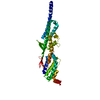


 PDBj
PDBj Assembly

 Mass: 18.015 Da / Num. of mol.: 221 / Source method: isolated from a natural source / Formula: H2O
 Sample preparation
 / Beamline: X25 / Wavelength: 1.04 Å
 Processing