 Movie
Movie Controller

[English] 日本語
 Yorodumi
- PDB-5a2u: Crystal structure of BBA68 or BbCRASP-1 from Borrelia burgdorferi... -
+ Open data

- Basic information
| Entry | Database: PDB / ID: 5a2u | ||||||
|---|---|---|---|---|---|---|---|
| Title | Crystal structure of BBA68 or BbCRASP-1 from Borrelia burgdorferi strain B31 | ||||||
 Components Components | COMPLEMENT REGULATOR ACQUIRING PROTEIN 1 | ||||||
 Keywords Keywords | IMMUNE SYSTEM / LYME DISEASE / LIPOPROTEIN / OUTER SURFACE PROTEIN | ||||||
| Function / homology | Bbcrasp-1 / Bbcrasp-1 / Borrelia lipoprotein paralogus family 54/60 / Borrelia Bbcrasp-1 domain containing protein / Orthogonal Bundle / Mainly Alpha / Complement regulator-acquiring surface protein 1 (CRASP-1) / Complement regulator acquiring protein 1 Function and homology information Function and homology information | ||||||
| Biological species |  BORRELIA BURGDORFERI (Lyme disease spirochete) BORRELIA BURGDORFERI (Lyme disease spirochete) | ||||||
| Method |  X-RAY DIFFRACTION / SYNCHROTRON / MOLECULAR REPLACEMENT / Resolution: 3.3 Å X-RAY DIFFRACTION / SYNCHROTRON / MOLECULAR REPLACEMENT / Resolution: 3.3 Å | ||||||
 Authors Authors | Brangulis, K. / Bertulis, E. / Petrovskis, I. / Kazaks, A. / Tars, K. | ||||||
 Citation Citation | Journal: To be Published Title: Crystal Structure of Bba68 or Bbcrasp-1 from Borrelia Burgdorferi Strain B31 Authors: Brangulis, K. / Petrovskis, I. / Kazaks, A. / Tars, K. | ||||||
| History |
|
- Structure visualization
| Structure viewer | Molecule: MolmilJmol/JSmol |
|---|
- Downloads & links
-Download
| PDBx/mmCIF format | 5a2u.cif.gz | 83.4 KB | Display | PDBx/mmCIF format |
|---|---|---|---|---|
| PDB format | pdb5a2u.ent.gz | 65.3 KB | Display | PDB format |
| PDBx/mmJSON format | 5a2u.json.gz | Tree view | PDBx/mmJSON format | |
| Others |  Other downloads Other downloads |
-Validation report
| Arichive directory | https://data.pdbj.org/pub/pdb/validation_reports/a2/5a2uftp://data.pdbj.org/pub/pdb/validation_reports/a2/5a2u | HTTPS FTP |
|---|
-Related structure data
| Related structure data |  1w33S S: Starting model for refinement |
|---|---|
| Similar structure data |


-Links
 PDBj
PDBj- Assembly
| Deposited unit | 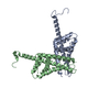
| ||||||||
|---|---|---|---|---|---|---|---|---|---|
| 1 | 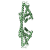
| ||||||||
| 2 | 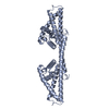
| ||||||||
| Unit cell |
|
-Components
| #1: Protein | Mass: 22575.879 Da / Num. of mol.: 2 / Fragment: UNP RESIDUES 65-251 Source method: isolated from a genetically manipulated source Source: (gene. exp.) BORRELIA BURGDORFERI (Lyme disease spirochete)Strain: B31 / Plasmid: PETM-11 / Production host: |
|---|
-Experimental details
-Experiment
| Experiment | Method: X-RAY DIFFRACTION / Number of used crystals: 1 |
|---|
- Sample preparation
| Crystal | Density Matthews: 2.9 Å3/Da / Density % sol: 58 % / Description: NONE |
|---|---|
| Crystal grow | pH: 8 / Details: 10% PEG 10 000 0.1M TRIS PH 8.0 |
-Data collection
| Diffraction | Mean temperature: 100 K |
|---|---|
| Diffraction source | Source: SYNCHROTRON / Site: MAX II  / Beamline: I911-3 / Wavelength: 1.0688 / Beamline: I911-3 / Wavelength: 1.0688 |
| Detector | Type: MARMOSAIC 225 mm CCD / Detector: CCD / Details: RH-COATED TOROIDAL SI MIRROR |
| Radiation | Monochromator: DOUBLE CRYSTAL SI(111) / Protocol: SINGLE WAVELENGTH / Monochromatic (M) / Laue (L): M / Scattering type: x-ray |
| Radiation wavelength | Wavelength: 1.0688 Å / Relative weight: 1 |
| Reflection | Resolution: 3.3→56.33 Å / Num. obs: 9488 / % possible obs: 96.9 % / Observed criterion σ(I): 2.4 / Redundancy: 4.2 % / Rmerge(I) obs: 0.04 / Net I/σ(I): 9.9 |
| Reflection shell | Resolution: 3.3→3.48 Å / Redundancy: 4.2 % / Rmerge(I) obs: 0.16 / Mean I/σ(I) obs: 3.3 / % possible all: 98.5 |
- Processing
| Software |
| ||||||||||||||||||||||||||||||||||||||||||||||||||||||||||||||||||||||||||||||||||||||||||||||||||||||||||||||||||||||||||||||||||||||||||||||||||||||||||||||||||||||||||||||||||||||
|---|---|---|---|---|---|---|---|---|---|---|---|---|---|---|---|---|---|---|---|---|---|---|---|---|---|---|---|---|---|---|---|---|---|---|---|---|---|---|---|---|---|---|---|---|---|---|---|---|---|---|---|---|---|---|---|---|---|---|---|---|---|---|---|---|---|---|---|---|---|---|---|---|---|---|---|---|---|---|---|---|---|---|---|---|---|---|---|---|---|---|---|---|---|---|---|---|---|---|---|---|---|---|---|---|---|---|---|---|---|---|---|---|---|---|---|---|---|---|---|---|---|---|---|---|---|---|---|---|---|---|---|---|---|---|---|---|---|---|---|---|---|---|---|---|---|---|---|---|---|---|---|---|---|---|---|---|---|---|---|---|---|---|---|---|---|---|---|---|---|---|---|---|---|---|---|---|---|---|---|---|---|---|---|
| Refinement | Method to determine structure: MOLECULAR REPLACEMENT Starting model: PDB ENTRY 1W33 Resolution: 3.3→50.45 Å / Cor.coef. Fo:Fc: 0.923 / Cor.coef. Fo:Fc free: 0.919 / SU B: 22.749 / SU ML: 0.389 / Cross valid method: THROUGHOUT / ESU R Free: 0.119 / Stereochemistry target values: MAXIMUM LIKELIHOOD Details: HYDROGENS HAVE BEEN ADDED IN THE RIDING POSITIONS. U VALUES REFINED INDIVIDUALLY
| ||||||||||||||||||||||||||||||||||||||||||||||||||||||||||||||||||||||||||||||||||||||||||||||||||||||||||||||||||||||||||||||||||||||||||||||||||||||||||||||||||||||||||||||||||||||
| Solvent computation | Ion probe radii: 0.8 Å / Shrinkage radii: 0.8 Å / VDW probe radii: 1.2 Å / Solvent model: MASK | ||||||||||||||||||||||||||||||||||||||||||||||||||||||||||||||||||||||||||||||||||||||||||||||||||||||||||||||||||||||||||||||||||||||||||||||||||||||||||||||||||||||||||||||||||||||
| Displacement parameters | Biso mean: 101.414 Å2
| ||||||||||||||||||||||||||||||||||||||||||||||||||||||||||||||||||||||||||||||||||||||||||||||||||||||||||||||||||||||||||||||||||||||||||||||||||||||||||||||||||||||||||||||||||||||
| Refinement step | Cycle: LAST / Resolution: 3.3→50.45 Å
| ||||||||||||||||||||||||||||||||||||||||||||||||||||||||||||||||||||||||||||||||||||||||||||||||||||||||||||||||||||||||||||||||||||||||||||||||||||||||||||||||||||||||||||||||||||||
| Refine LS restraints |
|