TRANSPORT PROTEIN / Tip20p / vesicle tethering / Endoplasmic reticulum / ER-Golgi transport / Membrane / Phosphoprotein / Protein transport / Transport
Function / homology
Function and homology information
Dsl1/NZR complex / regulation of ER to Golgi vesicle-mediated transport / COPI-dependent Golgi-to-ER retrograde traffic / retrograde vesicle-mediated transport, Golgi to endoplasmic reticulum / endoplasmic reticulum to Golgi vesicle-mediated transport / autophagy / protein transport / endoplasmic reticulum membrane / endoplasmic reticulum / cytosol Similarity search - Function
Dsl1p vesicle tethering complex, Tip20p subunit, domain E / Dsl1p vesicle tethering complex, Tip20p subunit, domain C / Dsl1p vesicle tethering complex, Tip20p subunit, domain B / Dsl1p vesicle tethering complex, Tip20p subunit, domain A / Dsl1p vesicle tethering complex, Tip20p subunit, domain D / RINT-1/Tip20 / Protein transport protein Tip20, domain E / Protein transport protein Tip20, domain A / Protein transport protein Tip20, domain B / Protein transport protein Tip20, domain C ...Dsl1p vesicle tethering complex, Tip20p subunit, domain E / Dsl1p vesicle tethering complex, Tip20p subunit, domain C / Dsl1p vesicle tethering complex, Tip20p subunit, domain B / Dsl1p vesicle tethering complex, Tip20p subunit, domain A / Dsl1p vesicle tethering complex, Tip20p subunit, domain D / RINT-1/Tip20 / Protein transport protein Tip20, domain E / Protein transport protein Tip20, domain A / Protein transport protein Tip20, domain B / Protein transport protein Tip20, domain C / RINT-1/TIP-1 family / RINT1/TIP20 domain profile. / EXOC6/PINT-1/Sec15/Tip20, C-terminal, domain 2 / Methane Monooxygenase Hydroxylase; Chain G, domain 1 / Tetracycline Repressor; domain 2 / Helix non-globular / Methane Monooxygenase Hydroxylase; Chain G, domain 1 / Special / Arc Repressor Mutant, subunit A / Up-down Bundle / Orthogonal Bundle / Mainly Alpha Similarity search - Domain/homology
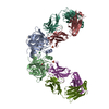
Mass: 82219.273 Da / Num. of mol.: 4 Source method: isolated from a genetically manipulated source Source: (gene. exp.) Saccharomyces cerevisiae (brewer's yeast) Gene: TIP20, TIP1, YGL145W / Production host: Escherichia coli (E. coli) / References: UniProt: P33891
Has protein modification
Y
-
Experimental details
-
Experiment
Experiment
Method: X-RAY DIFFRACTION / Number of used crystals: 1
-
Sample preparation
Crystal
Density Matthews: 3.99 Å3/Da / Density % sol: 69.14 %
Crystal grow
Temperature: 293 K / Method: vapor diffusion, hanging drop / pH: 6 Details: 3:1 ratio of protein (2 mg ml-1) and well buffer (0.1 M ADA, pH 6.0, 10% (w/v) PEG monomethyl ether 5K, 0.2 M LiSO4, 3% (v/v) isopropanol, 5 mM DTT), VAPOR DIFFUSION, HANGING DROP, temperature 293K
In the structure databanks used in Yorodumi, some data are registered as the other names, "COVID-19 virus" and "2019-nCoV". Here are the details of the virus and the list of structure data.
Jan 31, 2019. EMDB accession codes are about to change! (news from PDBe EMDB page)
EMDB accession codes are about to change! (news from PDBe EMDB page)
The allocation of 4 digits for EMDB accession codes will soon come to an end. Whilst these codes will remain in use, new EMDB accession codes will include an additional digit and will expand incrementally as the available range of codes is exhausted. The current 4-digit format prefixed with “EMD-” (i.e. EMD-XXXX) will advance to a 5-digit format (i.e. EMD-XXXXX), and so on. It is currently estimated that the 4-digit codes will be depleted around Spring 2019, at which point the 5-digit format will come into force.
The EM Navigator/Yorodumi systems omit the EMD- prefix.
Related info.:Q: What is EMD? / ID/Accession-code notation in Yorodumi/EM Navigator
Yorodumi is a browser for structure data from EMDB, PDB, SASBDB, etc.
This page is also the successor to EM Navigator detail page, and also detail information page/front-end page for Omokage search.
The word "yorodu" (or yorozu) is an old Japanese word meaning "ten thousand". "mi" (miru) is to see.
Related info.:EMDB / PDB / SASBDB / Comparison of 3 databanks / Yorodumi Search / Aug 31, 2016. New EM Navigator & Yorodumi / Yorodumi Papers / Jmol/JSmol / Function and homology information / Changes in new EM Navigator and Yorodumi
 Movie
Movie Controller


 Open data
 Basic information
 Components
 Keywords
 Function and homology information
 X-RAY DIFFRACTION /
  Authors
 Citation
 Structure visualization
 Downloads & links
 Other downloads
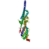


 PDBj
PDBj Assembly
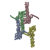


 Sample preparation
 / Beamline: X29A / Wavelength: 0.9792, 0.9794, 0.9640
 Processing