 Movie
Movie Controller

[English] 日本語
 Yorodumi
- PDB-1gh7: CRYSTAL STRUCTURE OF THE COMPLETE EXTRACELLULAR DOMAIN OF THE BET... -
+ Open data

- Basic information
| Entry | Database: PDB / ID: 1gh7 | |||||||||
|---|---|---|---|---|---|---|---|---|---|---|
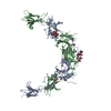
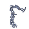
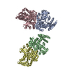
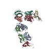
| Title | CRYSTAL STRUCTURE OF THE COMPLETE EXTRACELLULAR DOMAIN OF THE BETA-COMMON RECEPTOR OF IL-3, IL-5, AND GM-CSF | |||||||||
 Components Components | CYTOKINE RECEPTOR COMMON BETA CHAIN | |||||||||
 Keywords Keywords | CYTOKINE RECEPTOR / Dimer of interlocking chains of fibronectin-III domains Four fibronectin-III domains per chain | |||||||||
| Function / homology |  Function and homology information Function and homology informationDefective CSF2RB causes SMDP5 / Defective CSF2RA causes SMDP4 / granulocyte macrophage colony-stimulating factor receptor complex / granulocyte-macrophage colony-stimulating factor signaling pathway / respiratory gaseous exchange by respiratory system / interleukin-5-mediated signaling pathway / positive regulation of leukocyte proliferation / interleukin-3-mediated signaling pathway / cellular response to interleukin-3 / Surfactant metabolism ...Defective CSF2RB causes SMDP5 / Defective CSF2RA causes SMDP4 / granulocyte macrophage colony-stimulating factor receptor complex / granulocyte-macrophage colony-stimulating factor signaling pathway / respiratory gaseous exchange by respiratory system / interleukin-5-mediated signaling pathway / positive regulation of leukocyte proliferation / interleukin-3-mediated signaling pathway / cellular response to interleukin-3 / Surfactant metabolism / cytokine receptor activity / immunoglobulin mediated immune response / Interleukin-3, Interleukin-5 and GM-CSF signaling / Interleukin receptor SHC signaling / cell surface receptor signaling pathway via JAK-STAT / coreceptor activity / RAF/MAP kinase cascade / signaling receptor activity / response to lipopolysaccharide / external side of plasma membrane / signal transduction / plasma membrane Similarity search - Function | |||||||||
| Biological species |  Homo sapiens (human) Homo sapiens (human) | |||||||||
| Method |  X-RAY DIFFRACTION / SYNCHROTRON / Resolution: 3 Å X-RAY DIFFRACTION / SYNCHROTRON / Resolution: 3 Å | |||||||||
 Authors Authors | Carr, P.D. / Gustin, S.E. / Church, A.P. / Murphy, J.M. / Ford, S.C. / Mann, D.A. / Woltring, D.M. / Walker, I. / Ollis, D.L. / Young, I.G. | |||||||||
 Citation Citation | Journal: Cell(Cambridge,Mass.) / Year: 2001 Title: Structure of the complete extracellular domain of the common beta subunit of the human GM-CSF, IL-3, and IL-5 receptors reveals a novel dimer configuration. Authors: Carr, P.D. / Gustin, S.E. / Church, A.P. / Murphy, J.M. / Ford, S.C. / Mann, D.A. / Woltring, D.M. / Walker, I. / Ollis, D.L. / Young, I.G. | |||||||||
| History |
|
- Structure visualization
| Structure viewer | Molecule: MolmilJmol/JSmol |
|---|
- Downloads & links
-Download
| PDBx/mmCIF format | 1gh7.cif.gz | 173.4 KB | Display | PDBx/mmCIF format |
|---|---|---|---|---|
| PDB format | pdb1gh7.ent.gz | 136.3 KB | Display | PDB format |
| PDBx/mmJSON format | 1gh7.json.gz | Tree view | PDBx/mmJSON format | |
| Others |  Other downloads Other downloads |
-Validation report
| Arichive directory | https://data.pdbj.org/pub/pdb/validation_reports/gh/1gh7ftp://data.pdbj.org/pub/pdb/validation_reports/gh/1gh7 | HTTPS FTP |
|---|
-Related structure data
| Similar structure data |
|---|


-Links
 PDBj
PDBj


- Assembly
| Deposited unit | 
| ||||||||
|---|---|---|---|---|---|---|---|---|---|
| 1 |
| ||||||||
| Unit cell |
|
-Components
| #1: Protein | Mass: 47812.473 Da / Num. of mol.: 2 / Fragment: EXTRACELLULAR DOMAIN / Mutation: N328Q Source method: isolated from a genetically manipulated source Source: (gene. exp.) Homo sapiens (human)Cell line: CELL LINE HL60 DERIVED FROM A PROMYELOCYTIC LEUKEMIA Plasmid: PBACPAK8 (CLONTECH) Cell line (production host): BTI-TN-5BI-4 (HIGH FIVE) INVITROGEN Production host:  Trichoplusia ni (cabbage looper) / References: UniProt: P32927 Trichoplusia ni (cabbage looper) / References: UniProt: P32927#2: Polysaccharide | Source method: isolated from a genetically manipulated source #3: Polysaccharide | Source method: isolated from a genetically manipulated source Has protein modification | Y | |
|---|
-Experimental details
-Experiment
| Experiment | Method: X-RAY DIFFRACTION / Number of used crystals: 1 |
|---|
- Sample preparation
| Crystal | Density Matthews: 3.58 Å3/Da / Density % sol: 65.66 % |
|---|---|
| Crystal grow | Temperature: 277 K / Method: vapor diffusion, hanging drop / pH: 6.5 Details: PEG MME 5000, Phosphate, pH 6.5, VAPOR DIFFUSION/HANGING DROP, temperature 277K |
| Crystal grow | *PLUS Method: unknown |
-Data collection
| Diffraction | Mean temperature: 100 K |
|---|---|
| Diffraction source | Source: SYNCHROTRON / Site: ESRF  / Beamline: BM30A / Wavelength: 1.0085 / Beamline: BM30A / Wavelength: 1.0085 |
| Detector | Type: MAR scanner 345 mm plate / Detector: IMAGE PLATE / Date: Jul 18, 1999 |
| Radiation | Protocol: SINGLE WAVELENGTH / Monochromatic (M) / Laue (L): M / Scattering type: x-ray |
| Radiation wavelength | Wavelength: 1.0085 Å / Relative weight: 1 |
| Reflection | Resolution: 3→35 Å / Num. all: 281274 / Num. obs: 281274 / % possible obs: 99.8 % / Redundancy: 10.6 % / Rmerge(I) obs: 0.061 / Net I/σ(I): 20.3 |
| Reflection shell | Resolution: 3→3.11 Å / Rmerge(I) obs: 0.31 / Num. unique all: 2687 / % possible all: 100 |
| Reflection | *PLUS Num. obs: 26568 / Num. measured all: 281274 |
| Reflection shell | *PLUS % possible obs: 100 % / Num. unique obs: 2687 / Rmerge(I) obs: 0.31 |
- Processing
| Software |
| ||||||||||||||||||||
|---|---|---|---|---|---|---|---|---|---|---|---|---|---|---|---|---|---|---|---|---|---|
| Refinement | Resolution: 3→35 Å / Stereochemistry target values: Engh & Huber Details: Least squares minimization of amplitudes using X-PLOR followed by maximum likelihood refinement of amplitudes using CNS
| ||||||||||||||||||||
| Refinement step | Cycle: LAST / Resolution: 3→35 Å
| ||||||||||||||||||||
| Refine LS restraints |
| ||||||||||||||||||||
| Software | *PLUS Name: CNS / Classification: refinement | ||||||||||||||||||||
| Refinement | *PLUS Highest resolution: 3 Å / Lowest resolution: 35 Å / Rfactor obs: 0.267 / Rfactor Rfree: 0.304 | ||||||||||||||||||||
| Solvent computation | *PLUS | ||||||||||||||||||||
| Displacement parameters | *PLUS | ||||||||||||||||||||
| Refine LS restraints | *PLUS Type: c_bond_d / Dev ideal: 0.0092 |
