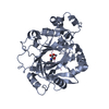
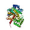
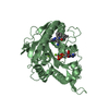
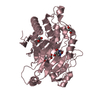
- PDB-3es1: Crystal structure of protein with a cupin-like fold and unknown f... -
+
Open data
ID or keywords:
Loading...
-
Basic information
Entry
Database: PDB / ID: 3es1
Title
Crystal structure of protein with a cupin-like fold and unknown function (YP_001165807.1) from NOVOSPHINGOBIUM AROMATICIVORANS DSM 12444 at 1.91 A resolution
Components
Cupin 2, conserved barrel domain protein
Keywords
structural genomics / unknown function / YP_001165807.1 / protein with a cupin-like fold and unknown function / Joint Center for Structural Genomics / JCSG / Protein Structure Initiative / PSI-2 / Cupin domain
Function / homology
Function and homology information
Ubiquitin Ligase Nedd4; Chain: W; - #150 / : / Ubiquitin Ligase Nedd4; Chain: W; / Cupin 2, conserved barrel / Cupin domain / RmlC-like cupin domain superfamily / Jelly Rolls / Single Sheet / RmlC-like jelly roll fold / Jelly Rolls ...Ubiquitin Ligase Nedd4; Chain: W; - #150 / : / Ubiquitin Ligase Nedd4; Chain: W; / Cupin 2, conserved barrel / Cupin domain / RmlC-like cupin domain superfamily / Jelly Rolls / Single Sheet / RmlC-like jelly roll fold / Jelly Rolls / Sandwich / Mainly Beta Similarity search - Domain/homology
Mass: 18.015 Da / Num. of mol.: 188 / Source method: isolated from a natural source / Formula: H2O
Has protein modification
Y
Sequence details
THE CONSTRUCT WAS EXPRESSED WITH A PURIFICATION TAG MGSDKIHHHHHHENLYFQG. THE TAG WAS REMOVED WITH ...THE CONSTRUCT WAS EXPRESSED WITH A PURIFICATION TAG MGSDKIHHHHHHENLYFQG. THE TAG WAS REMOVED WITH TEV PROTEASE LEAVING ONLY A GLYCINE (0) FOLLOWED BY THE TARGET SEQUENCE.
-
Experimental details
-
Experiment
Experiment
Method: X-RAY DIFFRACTION / Number of used crystals: 1
-
Sample preparation
Crystal
Density Matthews: 2.51 Å3/Da / Density % sol: 50.94 %
Crystal grow
Temperature: 277 K / Method: vapor diffusion, sitting drop / pH: 6.9 Details: 0.2M NaThioCyanate, 20.0% PEG-3350, No Buffer pH 6.9, NANODROP, VAPOR DIFFUSION, SITTING DROP, temperature 277K
Resolution: 1.91→28.665 Å / Num. obs: 15326 / % possible obs: 98 % / Observed criterion σ(I): -3 / Biso Wilson estimate: 22.05 Å2 / Rmerge(I) obs: 0.147 / Net I/σ(I): 13.43
Reflection shell
Resolution (Å)
Rmerge(I) obs
Mean I/σ(I) obs
Num. measured obs
Num. unique obs
Diffraction-ID
% possible all
1.91-1.98
0.011
2.1
22127
2830
1
97.4
1.98-2.06
0.011
2.9
22211
2828
1
97.6
2.06-2.15
0.011
4
20841
2666
1
97.5
2.15-2.26
0.011
4.8
21305
2725
1
97.8
2.26-2.41
0.011
5.9
23369
2972
1
97.9
2.41-2.59
0.011
7.5
21323
2711
1
98.2
2.59-2.85
0.011
10.7
22057
2808
1
98.4
2.85-3.26
0.011
18.2
22280
2832
1
98.5
3.26-4.1
0.011
33
22138
2828
1
98.9
4.1-28.665
0.011
44
22589
2872
1
98.2
-
Phasing
Phasing
Method: MAD
-
Processing
Software
Name
Version
Classification
NB
REFMAC
5.4.0067
refinement
PHENIX
refinement
SHELX
phasing
MolProbity
3beta29
modelbuilding
XSCALE
datascaling
PDB_EXTRACT
3.006
dataextraction
XDS
datareduction
SHARP
phasing
Refinement
Method to determine structure: MAD / Resolution: 1.91→28.665 Å / Cor.coef. Fo:Fc: 0.958 / Cor.coef. Fo:Fc free: 0.944 / Occupancy max: 1 / Occupancy min: 0.37 / SU B: 5.399 / SU ML: 0.087 / TLS residual ADP flag: LIKELY RESIDUAL / Cross valid method: THROUGHOUT / σ(F): 0 / ESU R: 0.136 / ESU R Free: 0.127 Stereochemistry target values: MAXIMUM LIKELIHOOD WITH PHASES Details: 1.HYDROGENS HAVE BEEN ADDED IN THE RIDING POSITIONS. 2.ATOM RECORD CONTAINS RESIDUAL B FACTORS ONLY. 3.A MET-INHIBITION PROTOCOL WAS USED FOR SELENOMETHIONINE INCORPORATION DURING PROTEIN ...Details: 1.HYDROGENS HAVE BEEN ADDED IN THE RIDING POSITIONS. 2.ATOM RECORD CONTAINS RESIDUAL B FACTORS ONLY. 3.A MET-INHIBITION PROTOCOL WAS USED FOR SELENOMETHIONINE INCORPORATION DURING PROTEIN EXPRESSION. THE OCCUPANCY OF THE SE ATOMS IN THE MSE RESIDUES WAS REDUCED TO 0.75 TO ACCOUNT FOR THE REDUCED SCATTERING POWER DUE TO PARTIAL S-MET INCORPORATION. 4.ONE CHLORIDE ION FROM CRYSTALLIZATION WAS MODELED IN THE STRUCTURE.
Rfactor
Num. reflection
% reflection
Selection details
Rfree
0.205
765
5 %
RANDOM
Rwork
0.17
-
-
-
obs
0.172
15326
97.54 %
-
Solvent computation
Ion probe radii: 0.8 Å / Shrinkage radii: 0.8 Å / VDW probe radii: 1.2 Å / Solvent model: MASK
In the structure databanks used in Yorodumi, some data are registered as the other names, "COVID-19 virus" and "2019-nCoV". Here are the details of the virus and the list of structure data.
Jan 31, 2019. EMDB accession codes are about to change! (news from PDBe EMDB page)
EMDB accession codes are about to change! (news from PDBe EMDB page)
The allocation of 4 digits for EMDB accession codes will soon come to an end. Whilst these codes will remain in use, new EMDB accession codes will include an additional digit and will expand incrementally as the available range of codes is exhausted. The current 4-digit format prefixed with “EMD-” (i.e. EMD-XXXX) will advance to a 5-digit format (i.e. EMD-XXXXX), and so on. It is currently estimated that the 4-digit codes will be depleted around Spring 2019, at which point the 5-digit format will come into force.
The EM Navigator/Yorodumi systems omit the EMD- prefix.
Related info.:Q: What is EMD? / ID/Accession-code notation in Yorodumi/EM Navigator
Yorodumi is a browser for structure data from EMDB, PDB, SASBDB, etc.
This page is also the successor to EM Navigator detail page, and also detail information page/front-end page for Omokage search.
The word "yorodu" (or yorozu) is an old Japanese word meaning "ten thousand". "mi" (miru) is to see.
Related info.:EMDB / PDB / SASBDB / Comparison of 3 databanks / Yorodumi Search / Aug 31, 2016. New EM Navigator & Yorodumi / Yorodumi Papers / Jmol/JSmol / Function and homology information / Changes in new EM Navigator and Yorodumi
 Movie
Movie Controller

 Yorodumi
 Open data
 Basic information
 Components
 Keywords
 Function and homology information
 Novosphingobium aromaticivorans DSM 12444 (bacteria)
 X-RAY DIFFRACTION /
  Authors
 Citation
 Structure visualization
 Downloads & links
 Other downloads
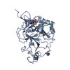


 PDBj
PDBj Assembly

 Mass: 35.453 Da / Num. of mol.: 1 / Source method: obtained synthetically / Formula: Cl
 Mass: 18.015 Da / Num. of mol.: 188 / Source method: isolated from a natural source / Formula: H2O
 Sample preparation
 / Beamline: BL9-2 / Wavelength: 0.92522,0.97932,0.97879
 Processing