- PDB-2gm1: Crystal structure of the mitotic kinesin eg5 in complex with mg-a... -
+
Open data
ID or keywords:
Loading...
-
Basic information
Entry
Database: PDB / ID: 2gm1
Title
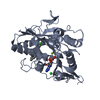
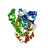
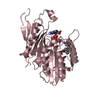
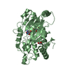
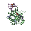
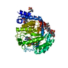
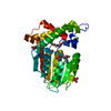
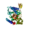
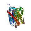
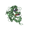
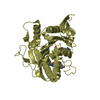
Crystal structure of the mitotic kinesin eg5 in complex with mg-adp and n-(3-aminopropyl)-n-((3-benzyl-5-chloro-4-oxo-3,4-dihydropyrrolo[2,1-f][1,2,4]triazin-2-yl)(cyclopropyl)methyl)-4-methylbenzamide
Components
KINESIN-RELATED MOTOR PROTEIN EG5
Keywords
CELL CYCLE / Eg5 Mg-ADP Complex Inhibitor
Function / homology
Function and homology information
spindle elongation / regulation of mitotic centrosome separation / plus-end-directed microtubule motor activity / mitotic centrosome separation / Kinesins / spindle organization / microtubule motor activity / kinesin complex / COPI-dependent Golgi-to-ER retrograde traffic / microtubule-based movement ...spindle elongation / regulation of mitotic centrosome separation / plus-end-directed microtubule motor activity / mitotic centrosome separation / Kinesins / spindle organization / microtubule motor activity / kinesin complex / COPI-dependent Golgi-to-ER retrograde traffic / microtubule-based movement / mitotic spindle assembly / MHC class II antigen presentation / mitotic spindle organization / sperm end piece / spindle / spindle pole / mitotic spindle / mitotic cell cycle / sperm principal piece / sperm midpiece / microtubule binding / microtubule / ciliary basal body / cell division / protein kinase binding / protein-containing complex / ATP binding / membrane / nucleus / cytosol Similarity search - Function
Kinesin-associated microtubule-binding domain / Kinesin-associated microtubule-binding / : / : / Kinesin motor domain / Kinesin / Kinesin motor domain signature. / Kinesin motor domain, conserved site / Kinesin motor domain / Kinesin motor domain profile. ...Kinesin-associated microtubule-binding domain / Kinesin-associated microtubule-binding / : / : / Kinesin motor domain / Kinesin / Kinesin motor domain signature. / Kinesin motor domain, conserved site / Kinesin motor domain / Kinesin motor domain profile. / Kinesin motor, catalytic domain. ATPase. / Kinesin motor domain / Kinesin motor domain superfamily / P-loop containing nucleoside triphosphate hydrolase / 3-Layer(aba) Sandwich / Alpha Beta Similarity search - Domain/homology
A: KINESIN-RELATED MOTOR PROTEIN EG5 B: KINESIN-RELATED MOTOR PROTEIN EG5 D: KINESIN-RELATED MOTOR PROTEIN EG5 E: KINESIN-RELATED MOTOR PROTEIN EG5 hetero molecules
Mass: 18.015 Da / Num. of mol.: 236 / Source method: isolated from a natural source / Formula: H2O
-
Experimental details
-
Experiment
Experiment
Method: X-RAY DIFFRACTION / Number of used crystals: 1
-
Sample preparation
Crystal
Density Matthews: 2.4 Å3/Da / Density % sol: 48.69 %
Crystal grow
Temperature: 277 K / pH: 7.5 Details: 1:1 reservoir: 0.1 M NaHEPES pH 7.5, 0.2 M MgCl2, 28% (v/v) PEG-3350; protein: 3.69 mg/ml (90 uM) in 20 mM PIPES pH 6.8, 2 mM MgCl2, 1 mM EGTA, 25 mM DTT, 478 uM ADP, 478 uM BMS-607205, 1.0% ...Details: 1:1 reservoir: 0.1 M NaHEPES pH 7.5, 0.2 M MgCl2, 28% (v/v) PEG-3350; protein: 3.69 mg/ml (90 uM) in 20 mM PIPES pH 6.8, 2 mM MgCl2, 1 mM EGTA, 25 mM DTT, 478 uM ADP, 478 uM BMS-607205, 1.0% DMSO , pH 7.8, vapor diffusion, hanging drop, temperature 277K
Protocol: SINGLE WAVELENGTH / Monochromatic (M) / Laue (L): M / Scattering type: x-ray
Radiation wavelength
Wavelength: 1 Å / Relative weight: 1
Reflection
Redundancy: 3.8 % / Av σ(I) over netI: 11.4 / Number: 254796 / Rmerge(I) obs: 0.067 / Χ2: 1.04 / D res high: 2.3 Å / D res low: 50 Å / Num. obs: 67500 / % possible obs: 99.7
Diffraction reflection shell
ID: 1
Highest resolution (Å)
Lowest resolution (Å)
Num. obs
% possible obs (%)
Rmerge(I) obs
Chi squared
Redundancy
4.95
50
6852
99.4
0.039
1.053
3.7
3.93
4.95
6809
100
0.042
0.989
3.8
3.44
3.93
6787
100
0.053
0.991
3.8
3.12
3.44
6771
100
0.066
1.147
3.8
2.9
3.12
6759
100
0.085
1.058
3.8
2.73
2.9
6724
100
0.111
1.056
3.8
2.59
2.73
6741
99.9
0.15
1.096
3.8
2.48
2.59
6700
99.5
0.184
1.028
3.7
2.38
2.48
6697
99.1
0.227
0.998
3.7
2.3
2.38
6660
99.1
0.271
0.958
3.7
Reflection
Resolution: 2.3→50 Å / Num. obs: 67500 / % possible obs: 99.7 % / Observed criterion σ(I): 0 / Redundancy: 3.8 % / Biso Wilson estimate: 24.6 Å2 / Rmerge(I) obs: 0.067 / Net I/σ(I): 18.2
Reflection shell
Resolution: 2.3→2.38 Å / Redundancy: 3.7 % / Rmerge(I) obs: 0.271 / Mean I/σ(I) obs: 5.1 / % possible all: 99.1
Method to determine structure: molecular replacement Starting model: EG5 TETRAMER Resolution: 2.3→49.86 Å / Rfactor Rfree error: 0.009 / Data cutoff high absF: 1956724.23 / Data cutoff low absF: 0 / Isotropic thermal model: RESTRAINED / Cross valid method: THROUGHOUT / σ(F): 0 / Stereochemistry target values: Engh & Huber Details: Due to a feature in the refinement program, the structure was refined with an OXT on a residue that is not the terminal residue of the sequence. The OXT was changed to N of the next residue ...Details: Due to a feature in the refinement program, the structure was refined with an OXT on a residue that is not the terminal residue of the sequence. The OXT was changed to N of the next residue by the wwPDB annotation staff.
In the structure databanks used in Yorodumi, some data are registered as the other names, "COVID-19 virus" and "2019-nCoV". Here are the details of the virus and the list of structure data.
Jan 31, 2019. EMDB accession codes are about to change! (news from PDBe EMDB page)
EMDB accession codes are about to change! (news from PDBe EMDB page)
The allocation of 4 digits for EMDB accession codes will soon come to an end. Whilst these codes will remain in use, new EMDB accession codes will include an additional digit and will expand incrementally as the available range of codes is exhausted. The current 4-digit format prefixed with “EMD-” (i.e. EMD-XXXX) will advance to a 5-digit format (i.e. EMD-XXXXX), and so on. It is currently estimated that the 4-digit codes will be depleted around Spring 2019, at which point the 5-digit format will come into force.
The EM Navigator/Yorodumi systems omit the EMD- prefix.
Related info.:Q: What is EMD? / ID/Accession-code notation in Yorodumi/EM Navigator
Yorodumi is a browser for structure data from EMDB, PDB, SASBDB, etc.
This page is also the successor to EM Navigator detail page, and also detail information page/front-end page for Omokage search.
The word "yorodu" (or yorozu) is an old Japanese word meaning "ten thousand". "mi" (miru) is to see.
Related info.:EMDB / PDB / SASBDB / Comparison of 3 databanks / Yorodumi Search / Aug 31, 2016. New EM Navigator & Yorodumi / Yorodumi Papers / Jmol/JSmol / Function and homology information / Changes in new EM Navigator and Yorodumi
 Movie
Movie Controller

 Yorodumi
 Open data
 Basic information
 Components
 Keywords
 Function and homology information
 Homo sapiens (human)
 X-RAY DIFFRACTION /
  Authors
 Citation
 Structure visualization
 Downloads & links
 Other downloads
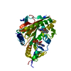
 PDBj
PDBj


 Assembly
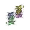


 Mass: 24.305 Da / Num. of mol.: 8 / Source method: obtained synthetically / Formula: Mg
 Mass: 504.023 Da / Num. of mol.: 4 / Source method: obtained synthetically / Formula: C28H30ClN5O2
 Mass: 427.201 Da / Num. of mol.: 4 / Source method: obtained synthetically / Formula: C10H15N5O10P2 / Comment: ADP, energy-carrying molecule*YM
 Mass: 18.015 Da / Num. of mol.: 236 / Source method: isolated from a natural source / Formula: H2O
 Sample preparation
 / Beamline: 17-ID / Wavelength: 1
  Processing