 Movie
Movie Controller


+ Open data

- Basic information
| Entry | Database: PDB / ID: 3els | ||||||
|---|---|---|---|---|---|---|---|
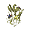
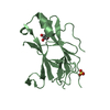
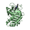
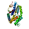
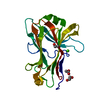
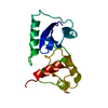
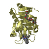
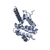
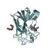
| Title | Crystal Structure of Yeast Pml1p, Residues 51-204 | ||||||
 Components Components | Pre-mRNA leakage protein 1 | ||||||
 Keywords Keywords | SPLICING / Intrinsically unstructured domain / forkhead-associated domain (FHA) domain / pre-mRNA retention and splicing / protein phosphorylation / RES complex / mRNA processing / mRNA splicing / Nucleus | ||||||
| Function / homology |  Function and homology information Function and homology informationmaintenance of RNA location / RES complex / U2-type prespliceosome assembly / U2 snRNP / mRNA export from nucleus / spliceosomal complex / mRNA splicing, via spliceosome / mRNA binding / nucleus / cytoplasm Similarity search - Function | ||||||
| Biological species |  | ||||||
| Method |  X-RAY DIFFRACTION / SYNCHROTRON / SAD / Resolution: 1.8 Å X-RAY DIFFRACTION / SYNCHROTRON / SAD / Resolution: 1.8 Å | ||||||
 Authors Authors | Trowitzsch, S. / Weber, G. / Luehrmann, R. / Wahl, M.C. | ||||||
 Citation Citation | Journal: J.Mol.Biol. / Year: 2009 Title: Crystal structure of the Pml1p subunit of the yeast precursor mRNA retention and splicing complex. Authors: Trowitzsch, S. / Weber, G. / Luhrmann, R. / Wahl, M.C. | ||||||
| History |
|
- Structure visualization
| Structure viewer | Molecule: MolmilJmol/JSmol |
|---|
- Downloads & links
-Download
| PDBx/mmCIF format | 3els.cif.gz | 50 KB | Display | PDBx/mmCIF format |
|---|---|---|---|---|
| PDB format | pdb3els.ent.gz | 35.1 KB | Display | PDB format |
| PDBx/mmJSON format | 3els.json.gz | Tree view | PDBx/mmJSON format | |
| Others |  Other downloads Other downloads |
-Validation report
| Arichive directory | https://data.pdbj.org/pub/pdb/validation_reports/el/3elsftp://data.pdbj.org/pub/pdb/validation_reports/el/3els | HTTPS FTP |
|---|
-Related structure data


-Links
 PDBj
PDBj

- Assembly
| Deposited unit | 
| ||||||||||||||||||||||||
|---|---|---|---|---|---|---|---|---|---|---|---|---|---|---|---|---|---|---|---|---|---|---|---|---|---|
| 1 |
| ||||||||||||||||||||||||
| 2 | x 6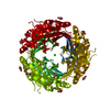
| ||||||||||||||||||||||||
| Unit cell |
| ||||||||||||||||||||||||
| Components on special symmetry positions |
|
-Components
| #1: Protein | Mass: 18050.314 Da / Num. of mol.: 1 / Fragment: UNP residues 51 to 204 Source method: isolated from a genetically manipulated source Source: (gene. exp.) Gene: PML1, YLR016C, L1591 / Plasmid: pCDFDuet-1 / Production host:  | ||
|---|---|---|---|
| #2: Chemical | ChemComp-MG /   Mass: 24.305 Da / Num. of mol.: 1 / Source method: obtained synthetically / Formula: Mg Mass: 24.305 Da / Num. of mol.: 1 / Source method: obtained synthetically / Formula: Mg | ||
| #3: Chemical | ChemComp-CL /   Mass: 35.453 Da / Num. of mol.: 1 / Source method: obtained synthetically / Formula: Cl Mass: 35.453 Da / Num. of mol.: 1 / Source method: obtained synthetically / Formula: Cl | ||
| #4: Chemical | ChemComp-GOL /   Mass: 92.094 Da / Num. of mol.: 4 / Source method: obtained synthetically / Formula: C3H8O3 Mass: 92.094 Da / Num. of mol.: 4 / Source method: obtained synthetically / Formula: C3H8O3#5: Water | ChemComp-HOH / |  Mass: 18.015 Da / Num. of mol.: 185 / Source method: isolated from a natural source / Formula: H2O Mass: 18.015 Da / Num. of mol.: 185 / Source method: isolated from a natural source / Formula: H2O |
-Experimental details
-Experiment
| Experiment | Method: X-RAY DIFFRACTION / Number of used crystals: 1 |
|---|
- Sample preparation
| Crystal | Density Matthews: 2.81 Å3/Da / Density % sol: 56.18 % |
|---|---|
| Crystal grow | Temperature: 293 K / Method: vapor diffusion, sitting drop / pH: 7.5 Details: 100 mM Tris-HCl, pH 7.5, 200 mM Li2SO4, 21-24 % PEG3350, VAPOR DIFFUSION, SITTING DROP, temperature 293K |
-Data collection
| Diffraction | Mean temperature: 100 K |
|---|---|
| Diffraction source | Source: SYNCHROTRON / Site: MPG/DESY, HAMBURG  / Beamline: BW6 / Wavelength: 0.9802 Å / Beamline: BW6 / Wavelength: 0.9802 Å |
| Detector | Type: MARMOSAIC 225 mm CCD / Detector: CCD / Date: Nov 1, 2007 / Details: mirrors |
| Radiation | Monochromator: Si 111 CHANNEL / Protocol: SINGLE WAVELENGTH / Monochromatic (M) / Laue (L): M / Scattering type: x-ray |
| Radiation wavelength | Wavelength: 0.9802 Å / Relative weight: 1 |
| Reflection | Resolution: 1.8→40.7 Å / Num. obs: 16896 / % possible obs: 99.9 % / Observed criterion σ(F): 0 / Observed criterion σ(I): 0 / Redundancy: 6.3 % / Biso Wilson estimate: 71.7 Å2 / Rsym value: 0.083 / Net I/σ(I): 19.7 |
| Reflection shell | Resolution: 2.4→2.53 Å / Redundancy: 6.2 % / Mean I/σ(I) obs: 1.6 / Num. unique all: 2473 / Rsym value: 0.81 / % possible all: 100 |
- Processing
| Software |
| ||||||||||||||||||||||||||||||||||||||||||||||||||||||||||||||||||||||||||||||||||||||||||
|---|---|---|---|---|---|---|---|---|---|---|---|---|---|---|---|---|---|---|---|---|---|---|---|---|---|---|---|---|---|---|---|---|---|---|---|---|---|---|---|---|---|---|---|---|---|---|---|---|---|---|---|---|---|---|---|---|---|---|---|---|---|---|---|---|---|---|---|---|---|---|---|---|---|---|---|---|---|---|---|---|---|---|---|---|---|---|---|---|---|---|---|
| Refinement | Method to determine structure: SAD / Resolution: 1.8→30 Å / Cor.coef. Fo:Fc: 0.968 / Cor.coef. Fo:Fc free: 0.945 / SU B: 2.859 / SU ML: 0.089 / Isotropic thermal model: Isotropic / Cross valid method: THROUGHOUT / σ(F): 0 / σ(I): 0 / ESU R: 0.12 / ESU R Free: 0.123 / Stereochemistry target values: MAXIMUM LIKELIHOOD / Details: HYDROGENS HAVE BEEN ADDED IN THE RIDING POSITIONS
| ||||||||||||||||||||||||||||||||||||||||||||||||||||||||||||||||||||||||||||||||||||||||||
| Solvent computation | Ion probe radii: 0.8 Å / Shrinkage radii: 0.8 Å / VDW probe radii: 1.4 Å / Solvent model: MASK | ||||||||||||||||||||||||||||||||||||||||||||||||||||||||||||||||||||||||||||||||||||||||||
| Displacement parameters | Biso mean: 36.309 Å2
| ||||||||||||||||||||||||||||||||||||||||||||||||||||||||||||||||||||||||||||||||||||||||||
| Refine analyze | Luzzati coordinate error obs: 0.21 Å | ||||||||||||||||||||||||||||||||||||||||||||||||||||||||||||||||||||||||||||||||||||||||||
| Refinement step | Cycle: LAST / Resolution: 1.8→30 Å
| ||||||||||||||||||||||||||||||||||||||||||||||||||||||||||||||||||||||||||||||||||||||||||
| Refine LS restraints |
| ||||||||||||||||||||||||||||||||||||||||||||||||||||||||||||||||||||||||||||||||||||||||||
| LS refinement shell | Resolution: 1.797→1.844 Å / Total num. of bins used: 20
|
