 Movie
Movie Controller

[English] 日本語
 Yorodumi
- PDB-2jkd: Structure of the yeast Pml1 splicing factor and its integration i... -
+ Open data

- Basic information
| Entry | Database: PDB / ID: 2jkd | ||||||
|---|---|---|---|---|---|---|---|
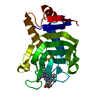
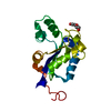
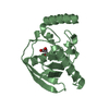
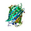
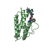
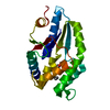
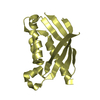
| Title | Structure of the yeast Pml1 splicing factor and its integration into the RES complex | ||||||
 Components Components | PRE-MRNA LEAKAGE PROTEIN 1 | ||||||
 Keywords Keywords | GENE REGULATION / MRNA PROCESSING / PML1/SNU17/BUD13 / PRE-MRNA SPLICING / SACCHAROMYCES CEREVISIAE / PHOSPHO-PEPTIDE RECOGNITION / RES / NUCLEUS / MRNA SPLICING | ||||||
| Function / homology |  Function and homology information Function and homology informationmaintenance of RNA location / RES complex / U2-type prespliceosome assembly / mRNA export from nucleus / mRNA splicing, via spliceosome / mRNA binding / nucleus / cytoplasm Similarity search - Function | ||||||
| Biological species |  | ||||||
| Method |  X-RAY DIFFRACTION / SYNCHROTRON / SAD / Resolution: 2.5 Å X-RAY DIFFRACTION / SYNCHROTRON / SAD / Resolution: 2.5 Å | ||||||
 Authors Authors | Brooks, M.A. / Dziembowski, A. / Quevillon-Cheruel, S. / Henriot, V. / Faux, C. / van Tilbeurgh, H. / Seraphin, B. | ||||||
 Citation Citation | Journal: Nucleic Acids Res. / Year: 2009 Title: Structure of the Yeast Pml1 Splicing Factor and its Integration Into the Res Complex Authors: Brooks, M.A. / Dziembowski, A. / Quevillon-Cheruel, S. / Henriot, V. / Faux, C. / Van Tilbeurgh, H. / Seraphin, B. | ||||||
| History |
|
- Structure visualization
| Structure viewer | Molecule: MolmilJmol/JSmol |
|---|
- Downloads & links
-Download
| PDBx/mmCIF format | 2jkd.cif.gz | 73 KB | Display | PDBx/mmCIF format |
|---|---|---|---|---|
| PDB format | pdb2jkd.ent.gz | 55 KB | Display | PDB format |
| PDBx/mmJSON format | 2jkd.json.gz | Tree view | PDBx/mmJSON format | |
| Others |  Other downloads Other downloads |
-Validation report
| Arichive directory | https://data.pdbj.org/pub/pdb/validation_reports/jk/2jkdftp://data.pdbj.org/pub/pdb/validation_reports/jk/2jkd | HTTPS FTP |
|---|
-Related structure data
| Similar structure data |
|---|


-Links
 PDBj
PDBj

- Assembly
| Deposited unit | 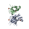
| ||||||||
|---|---|---|---|---|---|---|---|---|---|
| 1 | 
| ||||||||
| 2 | 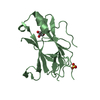
| ||||||||
| Unit cell |
| ||||||||
| Components on special symmetry positions |
| ||||||||
| Noncrystallographic symmetry (NCS) | NCS oper: (Code: given Matrix: (-0.671, -0.7413, 0.0138), Vector: |
-Components
| #1: Protein | Mass: 21543.215 Da / Num. of mol.: 2 / Fragment: FHA DOMAIN, RESIDUES 25-204 Source method: isolated from a genetically manipulated source Source: (gene. exp.) Strain: S288C / Plasmid: PET9 / Production host:  #2: Chemical |   Mass: 92.094 Da / Num. of mol.: 3 / Source method: obtained synthetically / Formula: C3H8O3 Mass: 92.094 Da / Num. of mol.: 3 / Source method: obtained synthetically / Formula: C3H8O3#3: Chemical |   Mass: 96.063 Da / Num. of mol.: 2 / Source method: obtained synthetically / Formula: SO4 Mass: 96.063 Da / Num. of mol.: 2 / Source method: obtained synthetically / Formula: SO4#4: Water | ChemComp-HOH / |  Mass: 18.015 Da / Num. of mol.: 46 / Source method: isolated from a natural source / Formula: H2O Mass: 18.015 Da / Num. of mol.: 46 / Source method: isolated from a natural source / Formula: H2OSequence details | RESIDUES 24-48 AND 113-121 OF CHAIN A AND 24-50 AND 112-121 OF CHAIN B WERE NOT MODELLED IN THE ...RESIDUES 24-48 AND 113-121 OF CHAIN A AND 24-50 AND 112-121 OF CHAIN B WERE NOT MODELLED IN THE STRUCTURE DUE TO LACK OF DENSITY. | |
|---|
-Experimental details
-Experiment
| Experiment | Method: X-RAY DIFFRACTION / Number of used crystals: 1 |
|---|
- Sample preparation
| Crystal | Density Matthews: 3.04 Å3/Da / Density % sol: 59.22 % / Description: NONE |
|---|---|
| Crystal grow | Temperature: 291 K / Method: vapor diffusion, hanging drop / pH: 8.5 Details: 0.1M TRIS-HCL, PH 8.5, 0.2 M LISO4 AND FROM 20 TO 30% PEG 4000 |
-Data collection
| Diffraction | Mean temperature: 100 K |
|---|---|
| Diffraction source | Source: SYNCHROTRON / Site: ESRF  / Beamline: ID14-4 / Wavelength: 0.979 / Beamline: ID14-4 / Wavelength: 0.979 |
| Detector | Type: ADSC CCD / Detector: CCD / Date: Aug 31, 2005 |
| Radiation | Protocol: SINGLE WAVELENGTH / Monochromatic (M) / Laue (L): M / Scattering type: x-ray |
| Radiation wavelength | Wavelength: 0.979 Å / Relative weight: 1 |
| Reflection | Resolution: 2.5→20 Å / Num. obs: 14550 / % possible obs: 98.2 % / Observed criterion σ(I): -3 / Redundancy: 2.94 % / Rmerge(I) obs: 0.038 / Net I/σ(I): 19.8 |
| Reflection shell | Resolution: 2.5→2.6 Å / Redundancy: 2.98 % / Rmerge(I) obs: 0.225 / Mean I/σ(I) obs: 5.82 / % possible all: 99.2 |
- Processing
| Software |
| ||||||||||||||||||||||||||||||||||||||||||||||||||||||||||||||||||||||||||||||||||||||||||||||||||||||||||||||||||||||||||||||||||||||||||||||||||||||||||||||||||||||||||||||||||||||
|---|---|---|---|---|---|---|---|---|---|---|---|---|---|---|---|---|---|---|---|---|---|---|---|---|---|---|---|---|---|---|---|---|---|---|---|---|---|---|---|---|---|---|---|---|---|---|---|---|---|---|---|---|---|---|---|---|---|---|---|---|---|---|---|---|---|---|---|---|---|---|---|---|---|---|---|---|---|---|---|---|---|---|---|---|---|---|---|---|---|---|---|---|---|---|---|---|---|---|---|---|---|---|---|---|---|---|---|---|---|---|---|---|---|---|---|---|---|---|---|---|---|---|---|---|---|---|---|---|---|---|---|---|---|---|---|---|---|---|---|---|---|---|---|---|---|---|---|---|---|---|---|---|---|---|---|---|---|---|---|---|---|---|---|---|---|---|---|---|---|---|---|---|---|---|---|---|---|---|---|---|---|---|---|
| Refinement | Method to determine structure: SAD Starting model: NONE Resolution: 2.5→40.86 Å / Cor.coef. Fo:Fc: 0.938 / Cor.coef. Fo:Fc free: 0.917 / SU B: 17.602 / SU ML: 0.191 / TLS residual ADP flag: LIKELY RESIDUAL / Cross valid method: THROUGHOUT / ESU R: 0.426 / ESU R Free: 0.281 / Stereochemistry target values: MAXIMUM LIKELIHOOD / Details: HYDROGENS HAVE BEEN ADDED IN THE RIDING POSITIONS.
| ||||||||||||||||||||||||||||||||||||||||||||||||||||||||||||||||||||||||||||||||||||||||||||||||||||||||||||||||||||||||||||||||||||||||||||||||||||||||||||||||||||||||||||||||||||||
| Solvent computation | Ion probe radii: 0.8 Å / Shrinkage radii: 0.8 Å / VDW probe radii: 1.4 Å / Solvent model: MASK | ||||||||||||||||||||||||||||||||||||||||||||||||||||||||||||||||||||||||||||||||||||||||||||||||||||||||||||||||||||||||||||||||||||||||||||||||||||||||||||||||||||||||||||||||||||||
| Displacement parameters | Biso mean: 50.84 Å2
| ||||||||||||||||||||||||||||||||||||||||||||||||||||||||||||||||||||||||||||||||||||||||||||||||||||||||||||||||||||||||||||||||||||||||||||||||||||||||||||||||||||||||||||||||||||||
| Refinement step | Cycle: LAST / Resolution: 2.5→40.86 Å
| ||||||||||||||||||||||||||||||||||||||||||||||||||||||||||||||||||||||||||||||||||||||||||||||||||||||||||||||||||||||||||||||||||||||||||||||||||||||||||||||||||||||||||||||||||||||
| Refine LS restraints |
|