 Movie
Movie Controller


+ Open data

- Basic information
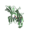
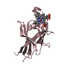
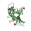
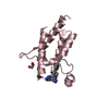
| Entry | Database: PDB / ID: 1pw6 | ||||||
|---|---|---|---|---|---|---|---|
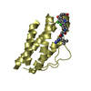
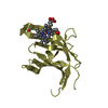
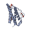
| Title | Low Micromolar Small Molecule Inhibitor of IL-2 | ||||||
 Components Components | Interleukin-2 | ||||||
 Keywords Keywords | IMMUNE SYSTEM / IL-2 interleukin 2 small molecule inhibitor | ||||||
| Function / homology |  Function and homology information Function and homology informationresponse to tacrolimus / kappa-type opioid receptor binding / regulation of CD4-positive, alpha-beta T cell proliferation / regulation of T cell homeostatic proliferation / interleukin-2 receptor binding / negative regulation of lymphocyte proliferation / glycosphingolipid binding / positive regulation of plasma cell differentiation / positive regulation of tissue remodeling / negative regulation of T-helper 17 cell differentiation ...response to tacrolimus / kappa-type opioid receptor binding / regulation of CD4-positive, alpha-beta T cell proliferation / regulation of T cell homeostatic proliferation / interleukin-2 receptor binding / negative regulation of lymphocyte proliferation / glycosphingolipid binding / positive regulation of plasma cell differentiation / positive regulation of tissue remodeling / negative regulation of T-helper 17 cell differentiation / RUNX1 and FOXP3 control the development of regulatory T lymphocytes (Tregs) / leukocyte activation involved in immune response / positive regulation of isotype switching to IgG isotypes / activated T cell proliferation / natural killer cell activation / Interleukin-2 signaling / cell surface receptor signaling pathway via STAT / kinase activator activity / positive regulation of activated T cell proliferation / positive regulation of regulatory T cell differentiation / negative regulation of B cell apoptotic process / interleukin-2-mediated signaling pathway / positive regulation of dendritic spine development / positive regulation of immunoglobulin production / positive regulation of interleukin-17 production / T cell differentiation / Interleukin receptor SHC signaling / extrinsic apoptotic signaling pathway in absence of ligand / positive regulation of B cell proliferation / cytokine activity / growth factor activity / negative regulation of inflammatory response / positive regulation of type II interferon production / positive regulation of inflammatory response / cell-cell signaling / positive regulation of cytosolic calcium ion concentration / carbohydrate binding / positive regulation of cell growth / RAF/MAP kinase cascade / transcription by RNA polymerase II / phospholipase C-activating G protein-coupled receptor signaling pathway / response to ethanol / adaptive immune response / cell adhesion / immune response / positive regulation of cell population proliferation / negative regulation of apoptotic process / positive regulation of transcription by RNA polymerase II / : / extracellular region Similarity search - Function | ||||||
| Biological species |  Homo sapiens (human) Homo sapiens (human) | ||||||
| Method |  X-RAY DIFFRACTION / MOLECULAR REPLACEMENT / Resolution: 2.6 Å X-RAY DIFFRACTION / MOLECULAR REPLACEMENT / Resolution: 2.6 Å | ||||||
 Authors Authors | Thanos, C.D. / Randal, M. / Wells, J.A. | ||||||
 Citation Citation | Journal: J.Am.Chem.Soc. / Year: 2003 Title: Potent small-molecule binding to a dynamic hot spot on IL-2. Authors: Thanos, C.D. / Randal, M. / Wells, J.A. | ||||||
| History |
|
- Structure visualization
| Structure viewer | Molecule: MolmilJmol/JSmol |
|---|
- Downloads & links
-Download
| PDBx/mmCIF format | 1pw6.cif.gz | 116.6 KB | Display | PDBx/mmCIF format |
|---|---|---|---|---|
| PDB format | pdb1pw6.ent.gz | 92 KB | Display | PDB format |
| PDBx/mmJSON format | 1pw6.json.gz | Tree view | PDBx/mmJSON format | |
| Others |  Other downloads Other downloads |
-Validation report
| Arichive directory | https://data.pdbj.org/pub/pdb/validation_reports/pw/1pw6ftp://data.pdbj.org/pub/pdb/validation_reports/pw/1pw6 | HTTPS FTP |
|---|
-Related structure data


-Links
 PDBj
PDBj


- Assembly
| Deposited unit | 
| ||||||||
|---|---|---|---|---|---|---|---|---|---|
| 1 | 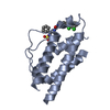
| ||||||||
| 2 | 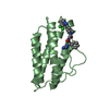
| ||||||||
| 3 | 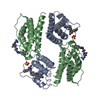
| ||||||||
| 4 | 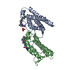
| ||||||||
| Unit cell |
|
-Components
| #1: Protein | Mass: 15435.979 Da / Num. of mol.: 2 Source method: isolated from a genetically manipulated source Source: (gene. exp.) Homo sapiens (human) / Gene: IL2 / Plasmid: prset / Species (production host): Escherichia coli / Production host:  #2: Chemical | ChemComp-SO4 / |   Mass: 96.063 Da / Num. of mol.: 1 / Source method: obtained synthetically / Formula: SO4 Mass: 96.063 Da / Num. of mol.: 1 / Source method: obtained synthetically / Formula: SO4#3: Chemical |   Mass: 534.481 Da / Num. of mol.: 2 / Source method: obtained synthetically / Formula: C25H33Cl2N7O2 Mass: 534.481 Da / Num. of mol.: 2 / Source method: obtained synthetically / Formula: C25H33Cl2N7O2Has protein modification | Y | |
|---|
-Experimental details
-Experiment
| Experiment | Method: X-RAY DIFFRACTION / Number of used crystals: 1 |
|---|
- Sample preparation
| Crystal | Density Matthews: 4.44 Å3/Da / Density % sol: 72.3 % | ||||||||||||||||||||||||
|---|---|---|---|---|---|---|---|---|---|---|---|---|---|---|---|---|---|---|---|---|---|---|---|---|---|
| Crystal grow | Method: vapor diffusion, hanging drop / pH: 5.9 Details: lithium sulfate, ammonium sulfate, pH 5.9, VAPOR DIFFUSION, HANGING DROP | ||||||||||||||||||||||||
| Crystal grow | *PLUS Method: vapor diffusion, hanging drop | ||||||||||||||||||||||||
| Components of the solutions | *PLUS
|
-Data collection
| Diffraction | Mean temperature: 93 K |
|---|---|
| Diffraction source | Source: ROTATING ANODE / Type: RIGAKU / Wavelength: 1.54 Å |
| Detector | Type: RIGAKU RAXIS IV / Detector: IMAGE PLATE / Date: Sep 9, 2001 |
| Radiation | Monochromator: Yale Mirrors / Protocol: SINGLE WAVELENGTH / Monochromatic (M) / Laue (L): M / Scattering type: x-ray |
| Radiation wavelength | Wavelength: 1.54 Å / Relative weight: 1 |
| Reflection | Resolution: 2.6→20 Å / Num. all: 17846 / Num. obs: 17310 / % possible obs: 97 % / Observed criterion σ(F): 0 / Observed criterion σ(I): 0 |
| Reflection shell | Resolution: 2.6→2.69 Å / % possible all: 97.9 |
| Reflection | *PLUS Highest resolution: 2.6 Å / Lowest resolution: 10 Å / Num. obs: 16176 / % possible obs: 97 % / Rmerge(I) obs: 0.143 |
| Reflection shell | *PLUS % possible obs: 97.9 % / Rmerge(I) obs: 0.374 / Mean I/σ(I) obs: 4.3 |
- Processing
| Software |
| |||||||||||||||||||||||||||||||||||||||||||||||||||||||||||||||||||||||||||
|---|---|---|---|---|---|---|---|---|---|---|---|---|---|---|---|---|---|---|---|---|---|---|---|---|---|---|---|---|---|---|---|---|---|---|---|---|---|---|---|---|---|---|---|---|---|---|---|---|---|---|---|---|---|---|---|---|---|---|---|---|---|---|---|---|---|---|---|---|---|---|---|---|---|---|---|---|
| Refinement | Method to determine structure: MOLECULAR REPLACEMENT / Resolution: 2.6→10 Å / Cor.coef. Fo:Fc: 0.859 / Cor.coef. Fo:Fc free: 0.824 / SU B: 14.087 / SU ML: 0.273 / Cross valid method: THROUGHOUT / σ(F): 0 / ESU R: 0.359 / ESU R Free: 0.282 / Stereochemistry target values: MAXIMUM LIKELIHOOD
| |||||||||||||||||||||||||||||||||||||||||||||||||||||||||||||||||||||||||||
| Solvent computation | Ion probe radii: 0.8 Å / Shrinkage radii: 0.8 Å / VDW probe radii: 1.4 Å / Solvent model: BABINET MODEL WITH MASK | |||||||||||||||||||||||||||||||||||||||||||||||||||||||||||||||||||||||||||
| Displacement parameters | Biso mean: 25.151 Å2
| |||||||||||||||||||||||||||||||||||||||||||||||||||||||||||||||||||||||||||
| Refinement step | Cycle: LAST / Resolution: 2.6→10 Å
| |||||||||||||||||||||||||||||||||||||||||||||||||||||||||||||||||||||||||||
| Refine LS restraints |
| |||||||||||||||||||||||||||||||||||||||||||||||||||||||||||||||||||||||||||
| LS refinement shell | Resolution: 2.6→2.685 Å / Total num. of bins used: 15 /
| |||||||||||||||||||||||||||||||||||||||||||||||||||||||||||||||||||||||||||
| Refinement TLS params. | Method: refined / Refine-ID: X-RAY DIFFRACTION
| |||||||||||||||||||||||||||||||||||||||||||||||||||||||||||||||||||||||||||
| Refinement TLS group |
| |||||||||||||||||||||||||||||||||||||||||||||||||||||||||||||||||||||||||||
| Refinement | *PLUS Lowest resolution: 10 Å / Rfactor Rfree: 0.292 / Rfactor Rwork: 0.257 | |||||||||||||||||||||||||||||||||||||||||||||||||||||||||||||||||||||||||||
| Solvent computation | *PLUS | |||||||||||||||||||||||||||||||||||||||||||||||||||||||||||||||||||||||||||
| Displacement parameters | *PLUS |
