 Movie
Movie Controller

[English] 日本語
 Yorodumi
- PDB-3egx: Crystal structure of the mammalian COPII-coat protein Sec23a/24a ... -
+ Open data

- Basic information
| Entry | Database: PDB / ID: 3egx | ||||||
|---|---|---|---|---|---|---|---|
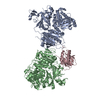
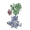
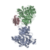
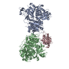
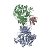
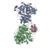
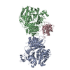
| Title | Crystal structure of the mammalian COPII-coat protein Sec23a/24a complexed with the SNARE protein Sec22b and bound to the transport signal sequence of the SNARE protein Bet1 | ||||||
 Components Components |
| ||||||
 Keywords Keywords | PROTEIN TRANSPORT / COPII coat protein / vesicle transport / transport signal sequence / Disease mutation / Endoplasmic reticulum / ER-Golgi transport / Golgi apparatus / Membrane / Transport / Phosphoprotein / Transmembrane | ||||||
| Function / homology |  Function and homology information Function and homology informationvesicle fusion with Golgi apparatus / regulation of cholesterol transport / COPII-coated vesicle cargo loading / COPII vesicle coat / SNARE complex / negative regulation of autophagosome assembly / SNAP receptor activity / Regulation of cholesterol biosynthesis by SREBP (SREBF) / cis-Golgi network / Cargo concentration in the ER ...vesicle fusion with Golgi apparatus / regulation of cholesterol transport / COPII-coated vesicle cargo loading / COPII vesicle coat / SNARE complex / negative regulation of autophagosome assembly / SNAP receptor activity / Regulation of cholesterol biosynthesis by SREBP (SREBF) / cis-Golgi network / Cargo concentration in the ER / retrograde vesicle-mediated transport, Golgi to endoplasmic reticulum / COPII-mediated vesicle transport / COPI-dependent Golgi-to-ER retrograde traffic / endoplasmic reticulum-Golgi intermediate compartment / endoplasmic reticulum exit site / endoplasmic reticulum to Golgi vesicle-mediated transport / transport vesicle / COPI-mediated anterograde transport / endoplasmic reticulum-Golgi intermediate compartment membrane / MHC class II antigen presentation / cholesterol homeostasis / GTPase activator activity / protein localization to plasma membrane / positive regulation of protein secretion / SNARE binding / Antigen Presentation: Folding, assembly and peptide loading of class I MHC / ER to Golgi transport vesicle membrane / intracellular protein transport / phagocytic vesicle membrane / positive regulation of protein catabolic process / melanosome / protein transport / ER-Phagosome pathway / Golgi membrane / endoplasmic reticulum membrane / perinuclear region of cytoplasm / SARS-CoV-2 activates/modulates innate and adaptive immune responses / endoplasmic reticulum / zinc ion binding / membrane / cytosol Similarity search - Function | ||||||
| Biological species |  Homo sapiens (human) Homo sapiens (human) | ||||||
| Method |  X-RAY DIFFRACTION / SYNCHROTRON / FOURIER SYNTHESIS / Resolution: 3.3 Å X-RAY DIFFRACTION / SYNCHROTRON / FOURIER SYNTHESIS / Resolution: 3.3 Å | ||||||
 Authors Authors | Goldberg, J. / Mancias, J.D. | ||||||
 Citation Citation | Journal: Embo J. / Year: 2008 Title: Structural basis of cargo membrane protein discrimination by the human COPII coat machinery. Authors: Mancias, J.D. / Goldberg, J. | ||||||
| History |
|
- Structure visualization
| Structure viewer | Molecule: MolmilJmol/JSmol |
|---|
- Downloads & links
-Download
| PDBx/mmCIF format | 3egx.cif.gz | 327.4 KB | Display | PDBx/mmCIF format |
|---|---|---|---|---|
| PDB format | pdb3egx.ent.gz | 258.1 KB | Display | PDB format |
| PDBx/mmJSON format | 3egx.json.gz | Tree view | PDBx/mmJSON format | |
| Others |  Other downloads Other downloads |
-Validation report
| Arichive directory | https://data.pdbj.org/pub/pdb/validation_reports/eg/3egxftp://data.pdbj.org/pub/pdb/validation_reports/eg/3egx | HTTPS FTP |
|---|
-Related structure data


-Links
 PDBj
PDBj


- Assembly
| Deposited unit | 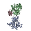
| ||||||||
|---|---|---|---|---|---|---|---|---|---|
| 1 |
| ||||||||
| Unit cell |
|
-Components
| #1: Protein | Mass: 86178.414 Da / Num. of mol.: 1 Source method: isolated from a genetically manipulated source Source: (gene. exp.) Homo sapiens (human) / Gene: SEC23A / Production host:   Spodoptera frugiperda (fall armyworm) / References: UniProt: Q15436 Spodoptera frugiperda (fall armyworm) / References: UniProt: Q15436 |
|---|---|
| #2: Protein | Mass: 84336.820 Da / Num. of mol.: 1 / Fragment: Conserved core, UNP residues 346-1093 Source method: isolated from a genetically manipulated source Source: (gene. exp.) Homo sapiens (human) / Gene: SEC24A / Production host: Spodoptera frugiperda (fall armyworm) / References: UniProt: O95486 |
| #3: Protein | Mass: 18031.645 Da / Num. of mol.: 1 / Fragment: cytoplasmic domainn, UNP residues 1-157 Source method: isolated from a genetically manipulated source Source: (gene. exp.) Homo sapiens (human) / Gene: SEC22B, SEC22L1 / Production host:  |
| #4: Protein/peptide | Mass: 1000.984 Da / Num. of mol.: 1 / Source method: obtained synthetically / Details: synthetic 9-residue peptide / References: UniProt: O15155*PLUS |
| #5: Chemical |   Mass: 65.409 Da / Num. of mol.: 2 / Source method: obtained synthetically / Formula: Zn Mass: 65.409 Da / Num. of mol.: 2 / Source method: obtained synthetically / Formula: Zn |
-Experimental details
-Experiment
| Experiment | Method: X-RAY DIFFRACTION / Number of used crystals: 1 |
|---|
- Sample preparation
| Crystal | Density Matthews: 2.46 Å3/Da / Density % sol: 50.01 % |
|---|---|
| Crystal grow | Temperature: 277 K / Method: vapor diffusion, hanging drop / pH: 7.9 Details: 10% (w/v) PEG 4000, 0.1 M NaCl, 0.5 M sodium acetate, 50 mM Tris buffer, pH 7.9, VAPOR DIFFUSION, HANGING DROP, temperature 277K |
-Data collection
| Diffraction | Mean temperature: 200 K |
|---|---|
| Diffraction source | Source: SYNCHROTRON / Site: NSLS  / Beamline: X25 / Wavelength: 1 Å / Beamline: X25 / Wavelength: 1 Å |
| Detector | Type: ADSC QUANTUM 4 / Detector: CCD / Date: May 5, 2007 |
| Radiation | Protocol: SINGLE WAVELENGTH / Monochromatic (M) / Laue (L): M / Scattering type: x-ray |
| Radiation wavelength | Wavelength: 1 Å / Relative weight: 1 |
| Reflection | Resolution: 3.3→25 Å / Num. obs: 24079 / % possible obs: 92 % / Observed criterion σ(F): 1 / Redundancy: 2.7 % / Rmerge(I) obs: 0.111 / Net I/σ(I): 9.6 |
| Reflection shell | Resolution: 3.3→3.4 Å / Redundancy: 2.6 % / Rmerge(I) obs: 0.394 / Mean I/σ(I) obs: 2.2 / % possible all: 95.1 |
- Processing
| Software |
| ||||||||||||||||||
|---|---|---|---|---|---|---|---|---|---|---|---|---|---|---|---|---|---|---|---|
| Refinement | Method to determine structure: FOURIER SYNTHESIS / Resolution: 3.3→25 Å / Cross valid method: THROUGHOUT / σ(F): 1 / Stereochemistry target values: Engh & Huber
| ||||||||||||||||||
| Refinement step | Cycle: LAST / Resolution: 3.3→25 Å
|
