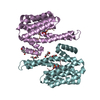
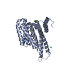
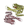
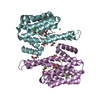
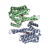
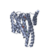
- PDB-3e6y: Structure of 14-3-3 in complex with the differentiation-inducing ... -
+
Open data
ID or keywords:
Loading...
-
Basic information
Entry
Database: PDB / ID: 3e6y
Title
Structure of 14-3-3 in complex with the differentiation-inducing agent Cotylenin A
Components
14-3-3-like protein C
H+-ATPase phosphopeptide QSYpTV
Keywords
SIGNALING PROTEIN / ADAPTER PROTEIN
Function / homology
Function and homology information
intracellular protein localization / signal transduction / ATP hydrolysis activity / ATP binding / membrane / metal ion binding / cytoplasm Similarity search - Function
Mass: 29399.977 Da / Num. of mol.: 2 Source method: isolated from a genetically manipulated source Source: (gene. exp.) Nicotiana tabacum (common tobacco) / Production host: Escherichia coli (E. coli) / References: UniProt: P93343
#2: Protein/peptide
H+-ATPasephosphopeptideQSYpTV
Mass: 676.610 Da / Num. of mol.: 2 / Source method: obtained synthetically Details: The peptide was chemically synthesized. It is found naturally in Arabidopsis thaliana References: UniProt: Q40409*PLUS
In the structure databanks used in Yorodumi, some data are registered as the other names, "COVID-19 virus" and "2019-nCoV". Here are the details of the virus and the list of structure data.
Jan 31, 2019. EMDB accession codes are about to change! (news from PDBe EMDB page)
EMDB accession codes are about to change! (news from PDBe EMDB page)
The allocation of 4 digits for EMDB accession codes will soon come to an end. Whilst these codes will remain in use, new EMDB accession codes will include an additional digit and will expand incrementally as the available range of codes is exhausted. The current 4-digit format prefixed with “EMD-” (i.e. EMD-XXXX) will advance to a 5-digit format (i.e. EMD-XXXXX), and so on. It is currently estimated that the 4-digit codes will be depleted around Spring 2019, at which point the 5-digit format will come into force.
The EM Navigator/Yorodumi systems omit the EMD- prefix.
Related info.:Q: What is EMD? / ID/Accession-code notation in Yorodumi/EM Navigator
Yorodumi is a browser for structure data from EMDB, PDB, SASBDB, etc.
This page is also the successor to EM Navigator detail page, and also detail information page/front-end page for Omokage search.
The word "yorodu" (or yorozu) is an old Japanese word meaning "ten thousand". "mi" (miru) is to see.
Related info.:EMDB / PDB / SASBDB / Comparison of 3 databanks / Yorodumi Search / Aug 31, 2016. New EM Navigator & Yorodumi / Yorodumi Papers / Jmol/JSmol / Function and homology information / Changes in new EM Navigator and Yorodumi
 Movie
Movie Controller

 Yorodumi
 Open data
 Basic information
 Components
 Keywords
 Function and homology information
 X-RAY DIFFRACTION /
  Authors
 Citation
 Structure visualization
 Downloads & links
 Other downloads
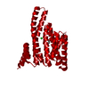


 PDBj
PDBj


 Assembly
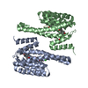


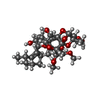
 Mass: 652.769 Da / Num. of mol.: 2 / Source method: obtained synthetically / Formula: C34H52O12
 Mass: 35.453 Da / Num. of mol.: 1 / Source method: obtained synthetically / Formula: Cl
 Mass: 18.015 Da / Num. of mol.: 100 / Source method: isolated from a natural source / Formula: H2O
 Sample preparation
 / Beamline: ID14-2 / Wavelength: 0.934 Å
 Processing