 Movie
Movie Controller

[English] 日本語
 Yorodumi
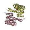
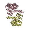
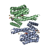
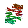
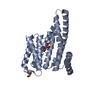
- PDB-1o9c: Structural view of a fungal toxin acting on a 14-3-3 regulatory c... -
+ Open data

- Basic information
| Entry | Database: PDB / ID: 1o9c | ||||||
|---|---|---|---|---|---|---|---|
| Title | Structural view of a fungal toxin acting on a 14-3-3 regulatory complex | ||||||
 Components Components | 14-3-3-LIKE PROTEIN C | ||||||
 Keywords Keywords | PROTEIN BINDING / FUSICOCCIN / 14-3-3 FAMILY / ACTIVATING DRUG / PLANT PLASMA MEMBRANE (H+)ATPASE | ||||||
| Function / homology |  Function and homology information Function and homology information | ||||||
| Biological species |  | ||||||
| Method |  X-RAY DIFFRACTION / SYNCHROTRON / MOLECULAR REPLACEMENT / Resolution: 2.6 Å X-RAY DIFFRACTION / SYNCHROTRON / MOLECULAR REPLACEMENT / Resolution: 2.6 Å | ||||||
 Authors Authors | Wurtele, M. / Jelich-Ottmann, C. / Wittinghofer, A. / Oecking, C. | ||||||
 Citation Citation | Journal: Embo J. / Year: 2003 Title: Structural View of a Fungal Toxin Acting on a 14-3-3 Regulatory Complex Authors: Wurtele, M. / Jelich-Ottmann, C. / Wittinghofer, A. / Oecking, C. | ||||||
| History |
|
- Structure visualization
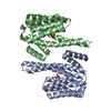
| Structure viewer | Molecule: MolmilJmol/JSmol |
|---|
- Downloads & links
-Download
| PDBx/mmCIF format | 1o9c.cif.gz | 59 KB | Display | PDBx/mmCIF format |
|---|---|---|---|---|
| PDB format | pdb1o9c.ent.gz | 43.1 KB | Display | PDB format |
| PDBx/mmJSON format | 1o9c.json.gz | Tree view | PDBx/mmJSON format | |
| Others |  Other downloads Other downloads |
-Validation report
| Arichive directory | https://data.pdbj.org/pub/pdb/validation_reports/o9/1o9cftp://data.pdbj.org/pub/pdb/validation_reports/o9/1o9c | HTTPS FTP |
|---|
-Related structure data
| Related structure data | 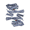 1o9dC 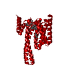 1o9eC 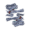 1o9fC 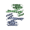 1a4oS C: citing same article ( S: Starting model for refinement |
|---|---|
| Similar structure data |


-Links
 PDBj
PDBj


- Assembly
| Deposited unit | 
| ||||||||
|---|---|---|---|---|---|---|---|---|---|
| 1 | 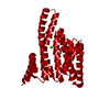
| ||||||||
| Unit cell |
|
-Components
| #1: Protein | Mass: 29399.977 Da / Num. of mol.: 1 Source method: isolated from a genetically manipulated source Details: GENE BANK AAC49892 / Source: (gene. exp.)  |
|---|---|
| #2: Chemical | ChemComp-FLC /   Mass: 189.100 Da / Num. of mol.: 1 / Source method: obtained synthetically / Formula: C6H5O7 Mass: 189.100 Da / Num. of mol.: 1 / Source method: obtained synthetically / Formula: C6H5O7 |
| #3: Chemical | ChemComp-CL /   Mass: 35.453 Da / Num. of mol.: 1 / Source method: obtained synthetically / Formula: Cl Mass: 35.453 Da / Num. of mol.: 1 / Source method: obtained synthetically / Formula: Cl |
| #4: Water | ChemComp-HOH /  Mass: 18.015 Da / Num. of mol.: 66 / Source method: isolated from a natural source / Formula: H2O Mass: 18.015 Da / Num. of mol.: 66 / Source method: isolated from a natural source / Formula: H2O |
-Experimental details
-Experiment
| Experiment | Method: X-RAY DIFFRACTION / Number of used crystals: 1 |
|---|
- Sample preparation
| Crystal | Density Matthews: 3.98 Å3/Da / Density % sol: 69.09 % | ||||||||||||||||||||||||||||||
|---|---|---|---|---|---|---|---|---|---|---|---|---|---|---|---|---|---|---|---|---|---|---|---|---|---|---|---|---|---|---|---|
| Crystal grow | pH: 6.4 / Details: PEG400, CITRATE PH 4.7, 0.2 MM AMMONIUM ACETATE | ||||||||||||||||||||||||||||||
| Crystal grow | *PLUS pH: 4.7 / Method: vapor diffusion, hanging drop | ||||||||||||||||||||||||||||||
| Components of the solutions | *PLUS
|
-Data collection
| Diffraction | Mean temperature: 100 K |
|---|---|
| Diffraction source | Source: SYNCHROTRON / Site: ESRF  / Beamline: ID29 / Wavelength: 0.8459 / Beamline: ID29 / Wavelength: 0.8459 |
| Detector | Date: Nov 1, 2001 |
| Radiation | Protocol: SINGLE WAVELENGTH / Monochromatic (M) / Laue (L): M / Scattering type: x-ray |
| Radiation wavelength | Wavelength: 0.8459 Å / Relative weight: 1 |
| Reflection | Resolution: 2.6→10 Å / Num. obs: 15163 / % possible obs: 99.1 % / Observed criterion σ(I): 2 / Redundancy: 13.6 % / Biso Wilson estimate: 44.4 Å2 / Rmerge(I) obs: 0.056 / Net I/σ(I): 33.1 |
| Reflection shell | Resolution: 2.6→2.7 Å / Rmerge(I) obs: 0.354 / Mean I/σ(I) obs: 8.2 / % possible all: 99.4 |
| Reflection | *PLUS Lowest resolution: 10 Å / Num. measured all: 205883 |
| Reflection shell | *PLUS % possible obs: 99.4 % |
- Processing
| Software |
| ||||||||||||||||||||||||||||||||||||||||||||||||||||||||||||
|---|---|---|---|---|---|---|---|---|---|---|---|---|---|---|---|---|---|---|---|---|---|---|---|---|---|---|---|---|---|---|---|---|---|---|---|---|---|---|---|---|---|---|---|---|---|---|---|---|---|---|---|---|---|---|---|---|---|---|---|---|---|
| Refinement | Method to determine structure: MOLECULAR REPLACEMENT Starting model: PDB ENTRY 1A4O Resolution: 2.6→19.75 Å / Rfactor Rfree error: 0.009 / Data cutoff high absF: 3345536.73 / Cross valid method: THROUGHOUT / σ(F): 0
| ||||||||||||||||||||||||||||||||||||||||||||||||||||||||||||
| Solvent computation | Solvent model: FLAT MODEL / Bsol: 37.8863 Å2 / ksol: 0.340786 e/Å3 | ||||||||||||||||||||||||||||||||||||||||||||||||||||||||||||
| Displacement parameters | Biso mean: 51.6 Å2
| ||||||||||||||||||||||||||||||||||||||||||||||||||||||||||||
| Refine analyze |
| ||||||||||||||||||||||||||||||||||||||||||||||||||||||||||||
| Refinement step | Cycle: LAST / Resolution: 2.6→19.75 Å
| ||||||||||||||||||||||||||||||||||||||||||||||||||||||||||||
| Refine LS restraints |
| ||||||||||||||||||||||||||||||||||||||||||||||||||||||||||||
| LS refinement shell | Resolution: 2.6→2.76 Å / Rfactor Rfree error: 0.032 / Total num. of bins used: 6
| ||||||||||||||||||||||||||||||||||||||||||||||||||||||||||||
| Refinement | *PLUS Highest resolution: 2.6 Å / Lowest resolution: 10 Å / % reflection Rfree: 5 % | ||||||||||||||||||||||||||||||||||||||||||||||||||||||||||||
| Solvent computation | *PLUS | ||||||||||||||||||||||||||||||||||||||||||||||||||||||||||||
| Displacement parameters | *PLUS | ||||||||||||||||||||||||||||||||||||||||||||||||||||||||||||
| Refine LS restraints | *PLUS
|
