SIGNALING PROTEIN / MULTIPLE SIGNALLING PATHWAYS / PHOSPHORYLATION / BINDING TO KINASE
Function / homology
Function and homology information
negative regulation of monoatomic ion transmembrane transport / small GTPase-mediated signal transduction / Regulation of localization of FOXO transcription factors / Activation of BAD and translocation to mitochondria / SARS-CoV-2 targets host intracellular signalling and regulatory pathways / SARS-CoV-1 targets host intracellular signalling and regulatory pathways / RHO GTPases activate PKNs / protein targeting / Chk1/Chk2(Cds1) mediated inactivation of Cyclin B:Cdk1 complex / 14-3-3 protein binding ...negative regulation of monoatomic ion transmembrane transport / small GTPase-mediated signal transduction / Regulation of localization of FOXO transcription factors / Activation of BAD and translocation to mitochondria / SARS-CoV-2 targets host intracellular signalling and regulatory pathways / SARS-CoV-1 targets host intracellular signalling and regulatory pathways / RHO GTPases activate PKNs / protein targeting / Chk1/Chk2(Cds1) mediated inactivation of Cyclin B:Cdk1 complex / 14-3-3 protein binding / substantia nigra development / TP53 Regulates Metabolic Genes / Translocation of SLC2A4 (GLUT4) to the plasma membrane / intracellular protein localization / transmembrane transporter binding / protein domain specific binding / focal adhesion / negative regulation of DNA-templated transcription / synapse / signal transduction / protein-containing complex / extracellular exosome / membrane / identical protein binding / nucleus / cytoplasm / cytosol Similarity search - Function
Mass: 18.015 Da / Num. of mol.: 169 / Source method: isolated from a natural source / Formula: H2O
-
Experimental details
-
Experiment
Experiment
Method: X-RAY DIFFRACTION / Number of used crystals: 1
-
Sample preparation
Crystal
Density Matthews: 2.3 Å3/Da / Density % sol: 46.9 % / Description: needle-like
Crystal grow
Temperature: 291 K / Method: vapor diffusion, hanging drop / pH: 6.4 Details: 24% PEG4K, 140 mM LI2SO4, 4 % of SATURATED NACL IN 80 mM MES AT pH 6.4; PROTEIN 7 mg/ml
In the structure databanks used in Yorodumi, some data are registered as the other names, "COVID-19 virus" and "2019-nCoV". Here are the details of the virus and the list of structure data.
Jan 31, 2019. EMDB accession codes are about to change! (news from PDBe EMDB page)
EMDB accession codes are about to change! (news from PDBe EMDB page)
The allocation of 4 digits for EMDB accession codes will soon come to an end. Whilst these codes will remain in use, new EMDB accession codes will include an additional digit and will expand incrementally as the available range of codes is exhausted. The current 4-digit format prefixed with “EMD-” (i.e. EMD-XXXX) will advance to a 5-digit format (i.e. EMD-XXXXX), and so on. It is currently estimated that the 4-digit codes will be depleted around Spring 2019, at which point the 5-digit format will come into force.
The EM Navigator/Yorodumi systems omit the EMD- prefix.
Related info.:Q: What is EMD? / ID/Accession-code notation in Yorodumi/EM Navigator
Yorodumi is a browser for structure data from EMDB, PDB, SASBDB, etc.
This page is also the successor to EM Navigator detail page, and also detail information page/front-end page for Omokage search.
The word "yorodu" (or yorozu) is an old Japanese word meaning "ten thousand". "mi" (miru) is to see.
Related info.:EMDB / PDB / SASBDB / Comparison of 3 databanks / Yorodumi Search / Aug 31, 2016. New EM Navigator & Yorodumi / Yorodumi Papers / Jmol/JSmol / Function and homology information / Changes in new EM Navigator and Yorodumi
 Movie
Movie Controller


 Open data
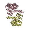
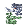
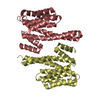
 Basic information
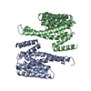
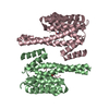
 Components
 Keywords
 Function and homology information
 Homo sapiens (human)
 X-RAY DIFFRACTION /
  Authors
 Citation
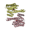
 Structure visualization
 Downloads & links
 Other downloads
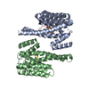


 PDBj
PDBj


 Assembly
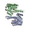


 Mass: 96.063 Da / Num. of mol.: 2 / Source method: obtained synthetically / Formula: SO4
 Mass: 18.015 Da / Num. of mol.: 169 / Source method: isolated from a natural source / Formula: H2O
 Sample preparation
 / Beamline: ID2 / Wavelength: 0.87 Å
 Processing