- PDB-3e38: CRYSTAL STRUCTURE OF A TWO-DOMAIN PROTEIN CONTAINING PREDICTED PH... -
+
Open data
ID or keywords:
Loading...
-
Basic information
Entry
Database: PDB / ID: 3.0E+38
Title
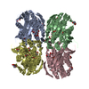
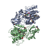
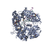
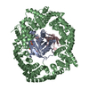
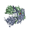
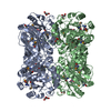
CRYSTAL STRUCTURE OF A TWO-DOMAIN PROTEIN CONTAINING PREDICTED PHP-LIKE METAL-DEPENDENT PHOSPHOESTERASE (BVU_3505) FROM BACTEROIDES VULGATUS ATCC 8482 AT 2.20 A RESOLUTION
Components
two-domain protein containing predicted PHP-like metal-dependent phosphoesterase
Keywords
HYDROLASE / Structural genomics / Joint Center for Structural Genomics / JCSG / Protein Structure Initiative / PSI-2
Function / homology
Function and homology information
5'-3' RNA exonuclease activity / 5'-3' DNA exonuclease activity / metal ion binding Similarity search - Function
Mass: 18.015 Da / Num. of mol.: 431 / Source method: isolated from a natural source / Formula: H2O
Has protein modification
Y
Sequence details
THE CONSTRUCT WAS EXPRESSED WITH A PURIFICATION TAG MGSDKIHHHHHHENLYFQG. THE TAG WAS REMOVED WITH ...THE CONSTRUCT WAS EXPRESSED WITH A PURIFICATION TAG MGSDKIHHHHHHENLYFQG. THE TAG WAS REMOVED WITH TEV PROTEASE LEAVING ONLY A GLYCINE (0) FOLLOWED BY THE TARGET SEQUENCE. THE CLONED CONSTRUCT CONTAINS RESIDUES 22-363 OF THE FULL LENGTH PROTEIN.
-
Experimental details
-
Experiment
Experiment
Method: X-RAY DIFFRACTION / Number of used crystals: 1
-
Sample preparation
Crystal
Density Matthews: 2.8 Å3/Da / Density % sol: 56.1 %
Crystal grow
Temperature: 277 K / Method: vapor diffusion, sitting drop / pH: 6.5 Details: 20.0000% Glycerol, 0.1600M Mg(oAc)2, 16.0000% PEG-8000, 0.1M Cacodylate pH 6.5, VAPOR DIFFUSION,SITTING DROP,NANODROP, temperature 277K, VAPOR DIFFUSION, SITTING DROP
Monochromator: Single crystal Si(111) bent monochromator (horizontal focusing) Protocol: SINGLE WAVELENGTH / Monochromatic (M) / Laue (L): M / Scattering type: x-ray
Radiation wavelength
Relative weight: 1
Reflection
Resolution: 2.2→29.604 Å / Num. obs: 45776 / % possible obs: 98.6 % / Observed criterion σ(I): -3 / Biso Wilson estimate: 31.645 Å2 / Rmerge(I) obs: 0.106 / Net I/σ(I): 11.94
Reflection shell
Resolution (Å)
Rmerge(I) obs
Mean I/σ(I) obs
Num. measured obs
Num. unique obs
Diffraction-ID
% possible all
2.2-2.28
0.77
2
28789
8350
1
93.2
2.28-2.37
0.622
2.4
31663
8579
1
99.2
2.37-2.48
0.496
3
33633
8962
1
99.3
2.48-2.61
0.393
3.8
32792
8689
1
99.4
2.61-2.77
0.31
4.8
32518
8561
1
99
2.77-2.98
0.214
6.8
33133
8654
1
99.4
2.98-3.28
0.129
10.8
33951
8845
1
99.5
3.28-3.76
0.074
18.2
33557
8850
1
99
3.76-4.72
0.039
29.8
33141
8686
1
99.2
4.72-29.604
0.031
37
34024
8827
1
98.5
-
Phasing
Phasing
Method: SAD
-
Processing
Software
Name
Version
Classification
NB
REFMAC
5.4.0067
refinement
PHENIX
refinement
SHELX
phasing
MolProbity
3beta29
modelbuilding
XSCALE
datascaling
PDB_EXTRACT
3.004
dataextraction
XDS
datareduction
SHELXD
phasing
autoSHARP
phasing
Refinement
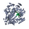
Method to determine structure: SAD / Resolution: 2.2→29.604 Å / Cor.coef. Fo:Fc: 0.961 / Cor.coef. Fo:Fc free: 0.935 / SU B: 10.417 / SU ML: 0.142 / TLS residual ADP flag: LIKELY RESIDUAL / Cross valid method: THROUGHOUT / σ(F): 0 / ESU R: 0.216 / ESU R Free: 0.188 Stereochemistry target values: MAXIMUM LIKELIHOOD WITH PHASES Details: 1.HYDROGENS HAVE BEEN ADDED IN THE RIDING POSITIONS. 2.ATOM RECORD CONTAINS RESIDUAL B FACTORS ONLY. 3.A MET-INHIBITION PROTOCOL WAS USED FOR SELENOMETHIONINE INCORPORATION DURING PROTEIN ...Details: 1.HYDROGENS HAVE BEEN ADDED IN THE RIDING POSITIONS. 2.ATOM RECORD CONTAINS RESIDUAL B FACTORS ONLY. 3.A MET-INHIBITION PROTOCOL WAS USED FOR SELENOMETHIONINE INCORPORATION DURING PROTEIN EXPRESSION. THE OCCUPANCY OF THE SE ATOMS IN THE MSE RESIDUES WAS REDUCED TO 0.75 TO ACCOUNT FOR THE REDUCED SCATTERING POWER DUE TO PARTIAL S-MET INCORPORATION. 4.GLYCEROL MOLECULES FROM THE CRYSTALLIZATION CONDITIONS ARE MODELED INTO THE STRUCTURE. 5. THREE ZN IONS, WHICH CO-PURIFIED WITH THE PROTEIN, ARE MODELED IN EACH SUBUNIT OF THIS STRUCTURE. ANOMALOUS DIFFERENCE FOURIERS AND X-RAY FLUORESCENCE EXPERIMENTS SUPPORT THE ASSIGNMENT OF THE ZINC ATOMS. 6.IN THE CONSERVED ACTIVE SITES, CACODYLATE ANIONS FROM THE CRYSTALLIZATION BUFFER HAVE BEEN MODELED. THE PRESENCE OF CACODYLATE AT THESE SITES IS SUPPORTED BY ANOMALOUS DIFFERENCE FOURIERS. HOWEVER, THE SITES ARE DISORDERED AND MAY ONLY BE PARTIALLY OCCUPIED. SINCE THIS DISORDER COULD NOT BE RESOLVED, ONLY A SINGLE ORIENTATION OF CACODYLATE IS MODELED IN THE STRUCTURE, EVEN THOUGH THIS DOES NOT FULLY SATISFY THE COORDINATION GEOMETRY OF THE ZN SITES.
Rfactor
Num. reflection
% reflection
Selection details
Rfree
0.225
2319
5.1 %
RANDOM
Rwork
0.172
-
-
-
obs
0.175
45753
98.77 %
-
Solvent computation
Ion probe radii: 0.8 Å / Shrinkage radii: 0.8 Å / VDW probe radii: 1.2 Å / Solvent model: MASK
In the structure databanks used in Yorodumi, some data are registered as the other names, "COVID-19 virus" and "2019-nCoV". Here are the details of the virus and the list of structure data.
Jan 31, 2019. EMDB accession codes are about to change! (news from PDBe EMDB page)
EMDB accession codes are about to change! (news from PDBe EMDB page)
The allocation of 4 digits for EMDB accession codes will soon come to an end. Whilst these codes will remain in use, new EMDB accession codes will include an additional digit and will expand incrementally as the available range of codes is exhausted. The current 4-digit format prefixed with “EMD-” (i.e. EMD-XXXX) will advance to a 5-digit format (i.e. EMD-XXXXX), and so on. It is currently estimated that the 4-digit codes will be depleted around Spring 2019, at which point the 5-digit format will come into force.
The EM Navigator/Yorodumi systems omit the EMD- prefix.
Related info.:Q: What is EMD? / ID/Accession-code notation in Yorodumi/EM Navigator
Yorodumi is a browser for structure data from EMDB, PDB, SASBDB, etc.
This page is also the successor to EM Navigator detail page, and also detail information page/front-end page for Omokage search.
The word "yorodu" (or yorozu) is an old Japanese word meaning "ten thousand". "mi" (miru) is to see.
Related info.:EMDB / PDB / SASBDB / Comparison of 3 databanks / Yorodumi Search / Aug 31, 2016. New EM Navigator & Yorodumi / Yorodumi Papers / Jmol/JSmol / Function and homology information / Changes in new EM Navigator and Yorodumi
 Movie
Movie Controller

 Yorodumi
 Open data
 Basic information
 Components
 Keywords
 Function and homology information
 Bacteroides vulgatus ATCC 8482 (bacteria)
 X-RAY DIFFRACTION /
  Authors
 Citation
 Structure visualization
 Downloads & links
 Other downloads
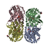

 PDBj
PDBj


 Assembly

 Mass: 65.409 Da / Num. of mol.: 6 / Source method: obtained synthetically / Formula: Zn
 Mass: 136.989 Da / Num. of mol.: 2 / Source method: obtained synthetically / Formula: C2H6AsO2
 Mass: 92.094 Da / Num. of mol.: 3 / Source method: obtained synthetically / Formula: C3H8O3
 Mass: 18.015 Da / Num. of mol.: 431 / Source method: isolated from a natural source / Formula: H2O
 Sample preparation
 / Beamline: BL11-1
 Processing