[English] 日本語
Yorodumi
- PDB-3l2n: Crystal structure of Putative carboxypeptidase A (YP_562911.1) fr... -

+
Open data


ID or keywords:

Loading...

-
Basic information

Entry
Database: PDB / ID: 3l2n
TitleCrystal structure of Putative carboxypeptidase A (YP_562911.1) from SHEWANELLA DENITRIFICANS OS-217 at 2.39 A resolution
ComponentsPeptidase M14, carboxypeptidase A
KeywordsHYDROLASE / Putative carboxypeptidase A / Structural Genomics / Joint Center for Structural Genomics / JCSG / Protein Structure Initiative / PSI-2 / Carboxypeptidase
Function / homology
Function and homology information


metallocarboxypeptidase activity / zinc ion binding
Similarity search - Function
Immunoglobulin-like - #3120 / Cytosolic carboxypeptidase, N-terminal / Cytosolic carboxypeptidase N-terminal domain / Zn_pept / Peptidase M14, carboxypeptidase A / Zinc carboxypeptidase / Zn peptidases / Aminopeptidase / Immunoglobulin-like / Sandwich ...Immunoglobulin-like - #3120 / Cytosolic carboxypeptidase, N-terminal / Cytosolic carboxypeptidase N-terminal domain / Zn_pept / Peptidase M14, carboxypeptidase A / Zinc carboxypeptidase / Zn peptidases / Aminopeptidase / Immunoglobulin-like / Sandwich / 3-Layer(aba) Sandwich / Mainly Beta / Alpha Beta
Similarity search - Domain/homology
ACETATE ION / Unknown ligand / Peptidase M14, carboxypeptidase A
Similarity search - Component
Biological speciesShewanella denitrificans (bacteria)
MethodX-RAY DIFFRACTION / SYNCHROTRON / SAD / Resolution: 2.39 Å
AuthorsJoint Center for Structural Genomics (JCSG)
CitationJournal: To be published
Title: Crystal structure of Putative carboxypeptidase A (YP_562911.1) from SHEWANELLA DENITRIFICANS OS-217 at 2.39 A resolution
Authors: Joint Center for Structural Genomics (JCSG)
History
DepositionDec 15, 2009Deposition site: RCSB / Processing site: RCSB
Revision 1.0Dec 29, 2009Provider: repository / Type: Initial release
Revision 1.1Jul 13, 2011Group: Advisory / Version format compliance
Revision 1.2Oct 25, 2017Group: Author supporting evidence / Refinement description / Category: pdbx_struct_assembly_auth_evidence / software
Revision 1.3Jul 17, 2019Group: Data collection / Derived calculations / Refinement description
Category: software / struct_conn
Item: _software.classification / _software.contact_author ..._software.classification / _software.contact_author / _software.contact_author_email / _software.language / _software.location / _software.name / _software.type / _software.version / _struct_conn.pdbx_leaving_atom_flag
Revision 1.4Feb 1, 2023Group: Database references / Derived calculations
Category: database_2 / pdbx_struct_conn_angle ...database_2 / pdbx_struct_conn_angle / struct_conn / struct_ref_seq_dif / struct_site
Item: _database_2.pdbx_DOI / _database_2.pdbx_database_accession ..._database_2.pdbx_DOI / _database_2.pdbx_database_accession / _pdbx_struct_conn_angle.ptnr1_auth_asym_id / _pdbx_struct_conn_angle.ptnr1_label_asym_id / _pdbx_struct_conn_angle.ptnr2_auth_asym_id / _pdbx_struct_conn_angle.ptnr2_label_asym_id / _pdbx_struct_conn_angle.ptnr3_auth_asym_id / _pdbx_struct_conn_angle.ptnr3_label_asym_id / _pdbx_struct_conn_angle.value / _struct_conn.conn_type_id / _struct_conn.id / _struct_conn.pdbx_dist_value / _struct_conn.pdbx_leaving_atom_flag / _struct_conn.ptnr1_auth_asym_id / _struct_conn.ptnr1_auth_comp_id / _struct_conn.ptnr1_auth_seq_id / _struct_conn.ptnr1_label_asym_id / _struct_conn.ptnr1_label_atom_id / _struct_conn.ptnr1_label_comp_id / _struct_conn.ptnr1_label_seq_id / _struct_conn.ptnr2_auth_asym_id / _struct_conn.ptnr2_auth_comp_id / _struct_conn.ptnr2_auth_seq_id / _struct_conn.ptnr2_label_asym_id / _struct_conn.ptnr2_label_atom_id / _struct_conn.ptnr2_label_comp_id / _struct_conn.ptnr2_label_seq_id / _struct_ref_seq_dif.details / _struct_site.pdbx_auth_asym_id / _struct_site.pdbx_auth_comp_id / _struct_site.pdbx_auth_seq_id

-
Structure visualization

Structure viewerMolecule:
MolmilJmol/JSmol

Downloads & links

-
Assembly

Deposited unit
A: Peptidase M14, carboxypeptidase A
B: Peptidase M14, carboxypeptidase A
hetero molecules


Theoretical massNumber of molelcules
Total (without water)89,8089
Polymers89,5172
Non-polymers2907
Water4,107228
1
A: Peptidase M14, carboxypeptidase A
B: Peptidase M14, carboxypeptidase A
hetero molecules

A: Peptidase M14, carboxypeptidase A
B: Peptidase M14, carboxypeptidase A
hetero molecules


Theoretical massNumber of molelcules
Total (without water)179,61518
Polymers179,0344
Non-polymers58114
Water724
TypeNameSymmetry operationNumber
identity operation1_555x,y,z1
crystal symmetry operation10_444-y-1,-x-1,-z-1/61
Buried area12380 Å2
ΔGint-73 kcal/mol
Surface area49360 Å2
MethodPISA
Unit cell
Length a, b, c (Å)81.417, 81.417, 479.685
Angle α, β, γ (deg.)90.000, 90.000, 120.000
Int Tables number178
Space group name H-MP6122
Noncrystallographic symmetry (NCS)NCS domain:
IDEns-IDDetails
11A
21B

NCS domain segments:
Dom-IDComponent-IDEns-IDRefine codeAuth asym-IDAuth seq-ID
1114A1 - 376
2114B1 - 376
Details
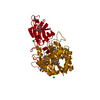
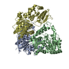
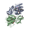
CRYSTAL PACKING ANALYSIS SUGGESTS THE ASSIGNMENT OF A TETRAMER AS THE SIGNIFICANT OLIGOMERIC FORM IN SOLUTION. ANALYTICAL SIZE-EXCLUSION CHROMATOGRAPHY COUPLED WITH STATIC LIGHT SCATTERING SUGGESTS A HEXAMER AS THE SOLUTION STATE OLIGOMERIC FORM.

-
Components

-
Protein , 1 types, 2 molecules AB

#1: Protein Peptidase M14, carboxypeptidase A


Mass: 44758.582 Da / Num. of mol.: 2
Source method: isolated from a genetically manipulated source
Source: (gene. exp.) Shewanella denitrificans (bacteria) / Strain: OS217 / ATCC BAA-1090 / DSM 15013 / Gene: Sden_1905 / Plasmid: SpeedET / Production host: Escherichia Coli (E. coli) / Strain (production host): HK100 / References: UniProt: Q12MY8

-
Non-polymers , 5 types, 235 molecules

#2: Chemical ChemComp-CA / CALCIUM ION


Mass: 40.078 Da / Num. of mol.: 2 / Source method: obtained synthetically / Formula: Ca
#3: Chemical ChemComp-UNL / UNKNOWN LIGAND


Num. of mol.: 2 / Source method: obtained synthetically
#4: Chemical ChemComp-ACT / ACETATE ION


Mass: 59.044 Da / Num. of mol.: 2 / Source method: obtained synthetically / Formula: C2H3O2
#5: Chemical ChemComp-GOL / GLYCEROL / GLYCERIN / PROPANE-1,2,3-TRIOL


Mass: 92.094 Da / Num. of mol.: 1 / Source method: obtained synthetically / Formula: C3H8O3
#6: Water ChemComp-HOH / water


Mass: 18.015 Da / Num. of mol.: 228 / Source method: isolated from a natural source / Formula: H2O

-
Details

Sequence detailsTHIS CONSTRUCT WAS EXPRESSED WITH A PURIFICATION TAG MGSDKIHHHHHHENLYFQG.

-
Experimental details

-
Experiment

ExperimentMethod: X-RAY DIFFRACTION / Number of used crystals: 1

-
Sample preparation

CrystalDensity Matthews: 2.56 Å3/Da / Density % sol: 52.02 %
Crystal growTemperature: 293 K / Method: vapor diffusion, sitting drop / pH: 5.6
Details: 15.0000% Glycerol, 0.1700M NH4OAc, 25.5000% PEG-4000, 0.1M Citrate pH 5.6, ADDITIVE: CALCIUM CHLORIDE 0.006 M, NANODROP', VAPOR DIFFUSION, SITTING DROP, temperature 293K

-
Data collection

DiffractionMean temperature: 100 K
Diffraction sourceSource: SYNCHROTRON / Site: SSRL / Beamline: BL11-1 / Wavelength: 0.97854
DetectorType: MARMOSAIC 325 mm CCD / Detector: CCD / Date: Jul 9, 2009 / Details: Flat mirror (vertical focusing)
RadiationMonochromator: Single crystal Si(111) bent monochromator (horizontal focusing)
Protocol: SINGLE WAVELENGTH / Monochromatic (M) / Laue (L): M / Scattering type: x-ray
Radiation wavelengthWavelength: 0.97854 Å / Relative weight: 1
ReflectionResolution: 2.39→49.147 Å / Num. obs: 38954 / % possible obs: 99.7 % / Observed criterion σ(I): -3 / Biso Wilson estimate: 40.987 Å2 / Rmerge(I) obs: 0.206 / Net I/σ(I): 13.16
Reflection shell
Resolution (Å)Rmerge(I) obsMean I/σ(I) obsNum. measured obsNum. unique obsDiffraction-ID% possible all
2.39-2.480.0132.5502923905198.9
2.48-2.570.0133.2490783418199.7
2.57-2.690.0133.7562203912199.8
2.69-2.830.0135539683768199.7
2.83-3.010.0136.4558523904199.7
3.01-3.240.0139.4540973801199.7
3.24-3.570.01314.55552639341100
3.57-4.080.01321.35475039201100
4.08-5.120.01329.65458439751100
5.12-49.1470.01331.7558634417199.5

-
Phasing

PhasingMethod: SAD

-
Processing

Software
NameVersionClassificationNB
REFMAC5.5.0053refinement
PHENIXrefinement
SHELXphasing
MolProbity3beta29model building
XSCALEdata scaling
PDB_EXTRACT3.006data extraction
XDSdata reduction
SHELXDphasing
autoSHARPphasing
RefinementMethod to determine structure: SAD / Resolution: 2.39→49.147 Å / Cor.coef. Fo:Fc: 0.953 / Cor.coef. Fo:Fc free: 0.922 / Occupancy max: 1 / Occupancy min: 0.5 / SU B: 15.453 / SU ML: 0.157 / TLS residual ADP flag: LIKELY RESIDUAL / Cross valid method: THROUGHOUT / σ(F): 0 / ESU R: 0.31 / ESU R Free: 0.231
Stereochemistry target values: MAXIMUM LIKELIHOOD WITH PHASES
Details: 1.HYDROGENS HAVE BEEN ADDED IN THE RIDING POSITIONS. 2.ATOM RECORDS CONTAIN RESIDUAL B FACTORS ONLY. 3.A MET-INHIBITION PROTOCOL WAS USED FOR SELENOMETHIONINE INCORPORATION DURING PROTEIN ...Details: 1.HYDROGENS HAVE BEEN ADDED IN THE RIDING POSITIONS. 2.ATOM RECORDS CONTAIN RESIDUAL B FACTORS ONLY. 3.A MET-INHIBITION PROTOCOL WAS USED FOR SELENOMETHIONINE INCORPORATION DURING PROTEIN EXPRESSION. THE OCCUPANCY OF THE SE ATOMS IN THE MSE RESIDUES WAS REDUCED TO 0.75 FOR THE REDUCED SCATTERING POWER DUE TO PARTIAL S-MET INCORPORATION. 4.CALCIUM (CA) HAS BEEN MODELED AT THE PUTATIVE ACTIVE SITE BASED ON ITS PRESENCE AS A CO-CRYSTALLIZATION COMPOUND AND A FLUORESCENCE BASED THERMAL SHIFT BINDING ASSAY. 5.AN UNIDENTIFIED LIGAND (UNL) RESEMBLING GLUTAMIC OR ASPARTIC ACID HAS BEEN MODELED AT THE PUTATIVE ACTIVE SITE. 6.ACETATE (ACT) AND GLYCEROL (GOL) FROM THE CRYSTALLIZATION SOLUTION HAVE BEEN MODELED IN THE SOLVENT STRUCTURE. 7.RAMACHANDRAN OUTLIERS AT RESIDUE 37 IN BOTH CHAINS ARE SUPPORTED BY ELECTRON DENSITY.
RfactorNum. reflection% reflectionSelection details
Rfree0.233 1947 5 %RANDOM
Rwork0.18 ---
obs0.182 38841 99.86 %-
Solvent computationIon probe radii: 0.8 Å / Shrinkage radii: 0.8 Å / VDW probe radii: 1.2 Å / Solvent model: MASK
Displacement parametersBiso max: 65.94 Å2 / Biso mean: 26.223 Å2 / Biso min: 3.25 Å2
Baniso -1Baniso -2Baniso -3
1-2.15 Å21.07 Å20 Å2
2--2.15 Å20 Å2
3----3.22 Å2
Refinement stepCycle: LAST / Resolution: 2.39→49.147 Å
ProteinNucleic acidLigandSolventTotal
Num. atoms5891 0 24 228 6143
Refine LS restraints
Refine-IDTypeDev idealDev ideal targetNumber
X-RAY DIFFRACTIONr_bond_refined_d0.0150.0226066
X-RAY DIFFRACTIONr_bond_other_d0.0010.023955
X-RAY DIFFRACTIONr_angle_refined_deg1.5461.9478271
X-RAY DIFFRACTIONr_angle_other_deg0.90739636
X-RAY DIFFRACTIONr_dihedral_angle_1_deg5.2815756
X-RAY DIFFRACTIONr_dihedral_angle_2_deg32.03124.901302
X-RAY DIFFRACTIONr_dihedral_angle_3_deg12.92415930
X-RAY DIFFRACTIONr_dihedral_angle_4_deg17.0261528
X-RAY DIFFRACTIONr_chiral_restr0.0870.2895
X-RAY DIFFRACTIONr_gen_planes_refined0.0060.0216921
X-RAY DIFFRACTIONr_gen_planes_other0.0010.021243
X-RAY DIFFRACTIONr_mcbond_it1.55833757
X-RAY DIFFRACTIONr_mcbond_other0.51631523
X-RAY DIFFRACTIONr_mcangle_it2.6556034
X-RAY DIFFRACTIONr_scbond_it5.26382309
X-RAY DIFFRACTIONr_scangle_it6.828112234
Refine LS restraints NCS

Dom-ID: 1 / Auth asym-ID: A / Ens-ID: 1 / Number: 4904 / Refine-ID: X-RAY DIFFRACTION

TypeRms dev position (Å)Weight position
MEDIUM POSITIONAL0.180.5
MEDIUM THERMAL0.942
LS refinement shellResolution: 2.39→2.452 Å / Total num. of bins used: 20
RfactorNum. reflection% reflection
Rfree0.331 141 -
Rwork0.271 2584 -
all-2725 -
obs--98.73 %
Refinement TLS params.

Method: refined / Refine-ID: X-RAY DIFFRACTION

IDL112)L122)L132)L222)L232)L332)S11 (Å °)S12 (Å °)S13 (Å °)S21 (Å °)S22 (Å °)S23 (Å °)S31 (Å °)S32 (Å °)S33 (Å °)T112)T122)T132)T222)T232)T332)Origin x (Å)Origin y (Å)Origin z (Å)
10.6927-0.1774-0.00450.67930.02220.8771-0.0158-0.0390.03780.0631-0.027-0.07530.00050.16410.04280.0567-0.0091-0.02040.05480.01280.0147-19.2629-29.2423-20.3076
20.7919-0.0395-0.04730.8025-0.36380.8012-0.0255-0.12130.10420.10750.05460.0254-0.1761-0.1305-0.02910.09610.04130.0090.0623-0.01560.0166-53.2867-16.1183-19.5685
Refinement TLS group
IDRefine-IDRefine TLS-IDAuth asym-IDAuth seq-ID
1X-RAY DIFFRACTION1A1 - 376
2X-RAY DIFFRACTION2B1 - 376

+
About Yorodumi

-
News

-
Feb 9, 2022. New format data for meta-information of EMDB entries

New format data for meta-information of EMDB entries

  • Version 3 of the EMDB header file is now the official format.
  • The previous official version 1.9 will be removed from the archive.

Related info.:EMDB header

External links:wwPDB to switch to version 3 of the EMDB data model

-
Aug 12, 2020. Covid-19 info

Covid-19 info

URL: https://pdbjlvh1.pdbj.org/emnavi/covid19.php

New page: Covid-19 featured information page in EM Navigator.

Related info.:Covid-19 info / Mar 5, 2020. Novel coronavirus structure data

+
Mar 5, 2020. Novel coronavirus structure data

Novel coronavirus structure data

Related info.:Yorodumi Speices / Aug 12, 2020. Covid-19 info

External links:COVID-19 featured content - PDBj / Molecule of the Month (242):Coronavirus Proteases

+
Jan 31, 2019. EMDB accession codes are about to change! (news from PDBe EMDB page)

EMDB accession codes are about to change! (news from PDBe EMDB page)

  • The allocation of 4 digits for EMDB accession codes will soon come to an end. Whilst these codes will remain in use, new EMDB accession codes will include an additional digit and will expand incrementally as the available range of codes is exhausted. The current 4-digit format prefixed with “EMD-” (i.e. EMD-XXXX) will advance to a 5-digit format (i.e. EMD-XXXXX), and so on. It is currently estimated that the 4-digit codes will be depleted around Spring 2019, at which point the 5-digit format will come into force.
  • The EM Navigator/Yorodumi systems omit the EMD- prefix.

Related info.:Q: What is EMD? / ID/Accession-code notation in Yorodumi/EM Navigator

External links:EMDB Accession Codes are Changing Soon! / Contact to PDBj

+
Jul 12, 2017. Major update of PDB

Major update of PDB

  • wwPDB released updated PDB data conforming to the new PDBx/mmCIF dictionary.
  • This is a major update changing the version number from 4 to 5, and with Remediation, in which all the entries are updated.
  • In this update, many items about electron microscopy experimental information are reorganized (e.g. em_software).
  • Now, EM Navigator and Yorodumi are based on the updated data.

External links:wwPDB Remediation / Enriched Model Files Conforming to OneDep Data Standards Now Available in the PDB FTP Archive

-
Yorodumi

Thousand views of thousand structures

  • Yorodumi is a browser for structure data from EMDB, PDB, SASBDB, etc.
  • This page is also the successor to EM Navigator detail page, and also detail information page/front-end page for Omokage search.
  • The word "yorodu" (or yorozu) is an old Japanese word meaning "ten thousand". "mi" (miru) is to see.

Related info.:EMDB / PDB / SASBDB / Comparison of 3 databanks / Yorodumi Search / Aug 31, 2016. New EM Navigator & Yorodumi / Yorodumi Papers / Jmol/JSmol / Function and homology information / Changes in new EM Navigator and Yorodumi

Read more