 Movie
Movie Controller


+ Open data

- Basic information
| Entry | Database: PDB / ID: 1q3g | ||||||
|---|---|---|---|---|---|---|---|
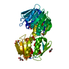
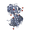
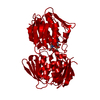
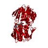
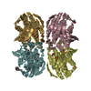
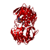
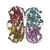
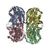
| Title | MurA (Asp305Ala) liganded with tetrahedral reaction intermediate | ||||||
 Components Components | UDP-N-acetylglucosamine 1-carboxyvinyltransferase | ||||||
 Keywords Keywords | TRANSFERASE / inside-out alpha-beta barrel | ||||||
| Function / homology |  Function and homology information Function and homology informationUDP-N-acetylglucosamine 1-carboxyvinyltransferase activity / UDP-N-acetylgalactosamine biosynthetic process / UDP-N-acetylglucosamine 1-carboxyvinyltransferase / peptidoglycan biosynthetic process / cell wall organization / regulation of cell shape / cell division / cytoplasm Similarity search - Function | ||||||
| Biological species |  Enterobacter cloacae (bacteria) Enterobacter cloacae (bacteria) | ||||||
| Method |  X-RAY DIFFRACTION / MOLECULAR REPLACEMENT / Resolution: 2.65 Å X-RAY DIFFRACTION / MOLECULAR REPLACEMENT / Resolution: 2.65 Å | ||||||
 Authors Authors | Eschenburg, S. / Kabsch, W. / Healy, M.L. / Schonbrunn, E. | ||||||
 Citation Citation | Journal: J.Biol.Chem. / Year: 2003 Title: A New View of the Mechanisms of UDP-N-Acetylglucosamine Enolpyruvyl Transferase (MurA) and 5-Enolpyruvylshikimate-3-phosphate Synthase (AroA) Derived from X-ray Structures of Their Tetrahedral ...Title: A New View of the Mechanisms of UDP-N-Acetylglucosamine Enolpyruvyl Transferase (MurA) and 5-Enolpyruvylshikimate-3-phosphate Synthase (AroA) Derived from X-ray Structures of Their Tetrahedral Reaction Intermediate States. Authors: Eschenburg, S. / Kabsch, W. / Healy, M.L. / Schonbrunn, E. | ||||||
| History |
|
- Structure visualization
| Structure viewer | Molecule: MolmilJmol/JSmol |
|---|
- Downloads & links
-Download
| PDBx/mmCIF format | 1q3g.cif.gz | 1.2 MB | Display | PDBx/mmCIF format |
|---|---|---|---|---|
| PDB format | pdb1q3g.ent.gz | 1 MB | Display | PDB format |
| PDBx/mmJSON format | 1q3g.json.gz | Tree view | PDBx/mmJSON format | |
| Others |  Other downloads Other downloads |
-Validation report
| Arichive directory | https://data.pdbj.org/pub/pdb/validation_reports/q3/1q3gftp://data.pdbj.org/pub/pdb/validation_reports/q3/1q3g | HTTPS FTP |
|---|
-Related structure data
| Related structure data |  1q36C 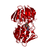 1a2nS S: Starting model for refinement C: citing same article ( |
|---|---|
| Similar structure data |

-Links
 PDBj
PDBj
- Assembly
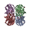
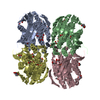
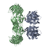
| Deposited unit | 
| ||||||||
|---|---|---|---|---|---|---|---|---|---|
| 1 | 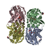
| ||||||||
| 2 | 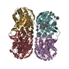
| ||||||||
| 3 | 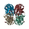
| ||||||||
| 4 | 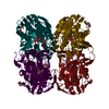
| ||||||||
| Unit cell |
|
-Components
| #1: Protein | Mass: 44785.398 Da / Num. of mol.: 16 / Mutation: N67D,D305A Source method: isolated from a genetically manipulated source Source: (gene. exp.) Enterobacter cloacae (bacteria) / Gene: MURA / Plasmid: pET9d / Species (production host): Escherichia coli / Production host: References: UniProt: P33038, UDP-N-acetylglucosamine 1-carboxyvinyltransferase #2: Chemical | ChemComp-UDA /   Mass: 775.396 Da / Num. of mol.: 16 / Source method: obtained synthetically / Formula: C20H32N3O23P3 Mass: 775.396 Da / Num. of mol.: 16 / Source method: obtained synthetically / Formula: C20H32N3O23P3#3: Chemical | ChemComp-EDO /   Mass: 62.068 Da / Num. of mol.: 16 / Source method: obtained synthetically / Formula: C2H6O2 Mass: 62.068 Da / Num. of mol.: 16 / Source method: obtained synthetically / Formula: C2H6O2#4: Water | ChemComp-HOH / |  Mass: 18.015 Da / Num. of mol.: 1431 / Source method: isolated from a natural source / Formula: H2O Mass: 18.015 Da / Num. of mol.: 1431 / Source method: isolated from a natural source / Formula: H2OHas protein modification | Y | |
|---|
-Experimental details
-Experiment
| Experiment | Method: X-RAY DIFFRACTION / Number of used crystals: 1 |
|---|
- Sample preparation
| Crystal | Density Matthews: 2.31 Å3/Da / Density % sol: 46.74 % | ||||||||||||||||||||||||||||||||||||
|---|---|---|---|---|---|---|---|---|---|---|---|---|---|---|---|---|---|---|---|---|---|---|---|---|---|---|---|---|---|---|---|---|---|---|---|---|---|
| Crystal grow | Temperature: 290 K / Method: vapor diffusion, hanging drop / pH: 6.4 Details: PEG 20,000, pH 6.4, VAPOR DIFFUSION, HANGING DROP, temperature 290K | ||||||||||||||||||||||||||||||||||||
| Crystal grow | *PLUS Temperature: 19 ℃ | ||||||||||||||||||||||||||||||||||||
| Components of the solutions | *PLUS
|
-Data collection
| Diffraction | Mean temperature: 90 K |
|---|---|
| Diffraction source | Source: ROTATING ANODE / Type: RIGAKU RU300 / Wavelength: 1.54 Å |
| Detector | Type: RIGAKU RAXIS IV / Detector: IMAGE PLATE / Details: mirrors |
| Radiation | Monochromator: osmic mirrors / Protocol: SINGLE WAVELENGTH / Monochromatic (M) / Laue (L): M / Scattering type: x-ray |
| Radiation wavelength | Wavelength: 1.54 Å / Relative weight: 1 |
| Reflection | Resolution: 2.65→20 Å / Num. all: 185835 / Num. obs: 185835 / % possible obs: 98.7 % / Observed criterion σ(F): 0 / Observed criterion σ(I): 0 / Redundancy: 4.4 % / Rmerge(I) obs: 0.098 / Net I/σ(I): 9.6 |
| Reflection shell | Resolution: 2.65→2.7 Å / Redundancy: 2.9 % / Rmerge(I) obs: 0.335 / Mean I/σ(I) obs: 2.3 / Num. unique all: 9008 / % possible all: 95.9 |
| Reflection | *PLUS Num. obs: 809759 / % possible obs: 98.9 % |
| Reflection shell | *PLUS Lowest resolution: 2.7 Å / % possible obs: 0.335 % / Num. unique obs: 26280 / Num. measured obs: 95.9 |
- Processing
| Software |
| ||||||||||||||||||||||||||||
|---|---|---|---|---|---|---|---|---|---|---|---|---|---|---|---|---|---|---|---|---|---|---|---|---|---|---|---|---|---|
| Refinement | Method to determine structure: MOLECULAR REPLACEMENT Starting model: PDB entry 1A2N Resolution: 2.65→20 Å / Cross valid method: THROUGHOUT / σ(F): 0 / Stereochemistry target values: Engh & Huber
| ||||||||||||||||||||||||||||
| Displacement parameters |
| ||||||||||||||||||||||||||||
| Refinement step | Cycle: LAST / Resolution: 2.65→20 Å
| ||||||||||||||||||||||||||||
| Refine LS restraints |
| ||||||||||||||||||||||||||||
| LS refinement shell | Resolution: 2.65→2.67 Å
| ||||||||||||||||||||||||||||
| Xplor file |
| ||||||||||||||||||||||||||||
| Refinement | *PLUS % reflection Rfree: 2 % / Rfactor Rwork: 0.216 | ||||||||||||||||||||||||||||
| Solvent computation | *PLUS | ||||||||||||||||||||||||||||
| Displacement parameters | *PLUS | ||||||||||||||||||||||||||||
| Refine LS restraints | *PLUS
| ||||||||||||||||||||||||||||
| LS refinement shell | *PLUS Lowest resolution: 2.7 Å |
