 Movie
Movie Controller


+ Open data

- Basic information
| Entry | Database: PDB / ID: 1naw | ||||||
|---|---|---|---|---|---|---|---|
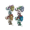
| Title | ENOLPYRUVYL TRANSFERASE | ||||||
 Components Components | UDP-N-ACETYLGLUCOSAMINE 1-CARBOXYVINYL-TRANSFERASE | ||||||
 Keywords Keywords | TRANSFERASE / PEPTIDOGLYCAN BIOSYNTHESIS / HINGE / DOMAIN MOVEMENT / SEQUENCE MOTIF / FOLDING | ||||||
| Function / homology |  Function and homology information Function and homology informationUDP-N-acetylglucosamine 1-carboxyvinyltransferase activity / UDP-N-acetylgalactosamine biosynthetic process / UDP-N-acetylglucosamine 1-carboxyvinyltransferase / peptidoglycan biosynthetic process / cell wall organization / regulation of cell shape / cell division / cytoplasm Similarity search - Function | ||||||
| Biological species |  Enterobacter cloacae (bacteria) Enterobacter cloacae (bacteria) | ||||||
| Method |  X-RAY DIFFRACTION / SYNCHROTRON / MIRAS / Resolution: 2 Å X-RAY DIFFRACTION / SYNCHROTRON / MIRAS / Resolution: 2 Å | ||||||
 Authors Authors | Schoenbrunn, E. / Sack, S. / Eschenburg, S. / Perrakis, A. / Krekel, F. / Amrhein, N. / Mandelkow, E. | ||||||
 Citation Citation | Journal: Structure / Year: 1996 Title: Crystal structure of UDP-N-acetylglucosamine enolpyruvyltransferase, the target of the antibiotic fosfomycin. Authors: Schonbrunn, E. / Sack, S. / Eschenburg, S. / Perrakis, A. / Krekel, F. / Amrhein, N. / Mandelkow, E. #1: Journal: J.Struct.Biol. / Year: 1996Title: Crystallization and Preliminary X-Ray Diffraction Analysis of Udp-N-Acetylglucosamine Enolpyruvyltransferase of Enterobacter Cloacae Authors: Sack, S. / Dauter, Z. / Wanke, C. / Amrhein, N. / Mandelkow, E. / Schonbrunn, E. #2: Journal: FEBS Lett. / Year: 1992Title: The Udp-N-Acetylglucosamine 1-Carboxyvinyl-Transferase of Enterobacter Cloacae. Molecular Cloning, Sequencing of the Gene and Overexpression of the Enzyme Authors: Wanke, C. / Falchetto, R. / Amrhein, N. | ||||||
| History |
|
- Structure visualization
| Structure viewer | Molecule: MolmilJmol/JSmol |
|---|
- Downloads & links
-Download
| PDBx/mmCIF format | 1naw.cif.gz | 179.2 KB | Display | PDBx/mmCIF format |
|---|---|---|---|---|
| PDB format | pdb1naw.ent.gz | 139.9 KB | Display | PDB format |
| PDBx/mmJSON format | 1naw.json.gz | Tree view | PDBx/mmJSON format | |
| Others |  Other downloads Other downloads |
-Validation report
| Arichive directory | https://data.pdbj.org/pub/pdb/validation_reports/na/1nawftp://data.pdbj.org/pub/pdb/validation_reports/na/1naw | HTTPS FTP |
|---|
-Related structure data
| Similar structure data |
|---|


-Links
 PDBj
PDBj
- Assembly
| Deposited unit | 
| |||||||||||||||
|---|---|---|---|---|---|---|---|---|---|---|---|---|---|---|---|---|
| 1 | 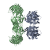
| |||||||||||||||
| 2 | 
| |||||||||||||||
| Unit cell |
| |||||||||||||||
| Components on special symmetry positions |
|
-Components
| #1: Protein | Mass: 44828.422 Da / Num. of mol.: 2 Source method: isolated from a genetically manipulated source Source: (gene. exp.) Enterobacter cloacae (bacteria) / Plasmid: PKK233-2 / Production host: References: UniProt: P33038, UDP-N-acetylglucosamine 1-carboxyvinyltransferase #2: Chemical |   Mass: 100.182 Da / Num. of mol.: 2 / Source method: obtained synthetically / Formula: C6H14N Mass: 100.182 Da / Num. of mol.: 2 / Source method: obtained synthetically / Formula: C6H14N#3: Water | ChemComp-HOH / |  Mass: 18.015 Da / Num. of mol.: 478 / Source method: isolated from a natural source / Formula: H2O Mass: 18.015 Da / Num. of mol.: 478 / Source method: isolated from a natural source / Formula: H2OCompound details | ENOLPYRUVY | |
|---|
-Experimental details
-Experiment
| Experiment | Method: X-RAY DIFFRACTION / Number of used crystals: 1 |
|---|
- Sample preparation
| Crystal | Density Matthews: 3.3 Å3/Da / Density % sol: 63 % | ||||||||||||||||||||
|---|---|---|---|---|---|---|---|---|---|---|---|---|---|---|---|---|---|---|---|---|---|
| Crystal grow | Method: vapor diffusion, hanging drop / pH: 6.4 Details: CRYSTALS WERE GROWN BY THE HANGING DROP VAPOR DIFFUSION METHOD USING PLASTIC TISSUE CULTURE PLATES. 0.4 M SODIUM/POTASSIUM PHOSPHATE BUFFER (PH 6.4) CONTAINING 40 MM CYCLOHEXYLAMMONIUM ...Details: CRYSTALS WERE GROWN BY THE HANGING DROP VAPOR DIFFUSION METHOD USING PLASTIC TISSUE CULTURE PLATES. 0.4 M SODIUM/POTASSIUM PHOSPHATE BUFFER (PH 6.4) CONTAINING 40 MM CYCLOHEXYLAMMONIUM PHOSPHATE WERE EQUILIBRATED AGAINST 1 ML 0.8 M SODIUM/POTASSIUM PHOSPHATE BUFFER (PH 6.4). CRYSTALLIZATION USUALLY OCCURRED WITHIN 3 DAYS AND THE CRYSTALS REACHED THEIR MAXIMUM SIZE OF 0.5 X 0.5 X 0.1 MM==3== AFTER 5 DAYS AT ROOM TEMPERATURE. (SEE REFERENCE 1 FOR DETAILS.), vapor diffusion - hanging drop Temp details: room temp | ||||||||||||||||||||
| Crystal grow | *PLUS Method: vapor diffusion, hanging drop | ||||||||||||||||||||
| Components of the solutions | *PLUS
|
-Data collection
| Diffraction | Mean temperature: 120 K |
|---|---|
| Diffraction source | Source: SYNCHROTRON / Site: EMBL/DESY, HAMBURG  / Beamline: X31 / Wavelength: 0.99 / Beamline: X31 / Wavelength: 0.99 |
| Detector | Type: MARRESEARCH / Detector: IMAGE PLATE / Date: Nov 27, 1995 |
| Radiation | Monochromator: DOUBLE CRYSTAL SI(111) / Monochromatic (M) / Laue (L): M / Scattering type: x-ray |
| Radiation wavelength | Wavelength: 0.99 Å / Relative weight: 1 |
| Reflection | Resolution: 1.8→25 Å / Num. obs: 103189 / % possible obs: 99.8 % / Redundancy: 7.2 % / Rmerge(I) obs: 0.053 |
| Reflection shell | Resolution: 1.8→1.84 Å / Redundancy: 3.3 % / Rmerge(I) obs: 0.29 / % possible all: 98.7 |
| Reflection | *PLUS Num. measured all: 744941 |
| Reflection shell | *PLUS % possible obs: 98.7 % |
- Processing
| Software |
| ||||||||||||||||||||||||||||||||||||||||||||||||||||||||||||||||||||||||||||||||||||
|---|---|---|---|---|---|---|---|---|---|---|---|---|---|---|---|---|---|---|---|---|---|---|---|---|---|---|---|---|---|---|---|---|---|---|---|---|---|---|---|---|---|---|---|---|---|---|---|---|---|---|---|---|---|---|---|---|---|---|---|---|---|---|---|---|---|---|---|---|---|---|---|---|---|---|---|---|---|---|---|---|---|---|---|---|---|
| Refinement | Method to determine structure: MIRAS / Resolution: 2→10 Å
| ||||||||||||||||||||||||||||||||||||||||||||||||||||||||||||||||||||||||||||||||||||
| Displacement parameters | Biso mean: 23.1 Å2 | ||||||||||||||||||||||||||||||||||||||||||||||||||||||||||||||||||||||||||||||||||||
| Refine analyze | Luzzati d res low obs: 10 Å / Luzzati sigma a obs: 0.24 Å | ||||||||||||||||||||||||||||||||||||||||||||||||||||||||||||||||||||||||||||||||||||
| Refinement step | Cycle: LAST / Resolution: 2→10 Å
| ||||||||||||||||||||||||||||||||||||||||||||||||||||||||||||||||||||||||||||||||||||
| Refine LS restraints |
|
