 Movie
Movie Controller


+ Open data

- Basic information
| Entry | Database: PDB / ID: 1vcb | ||||||
|---|---|---|---|---|---|---|---|
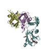
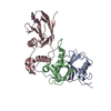
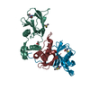
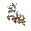
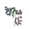
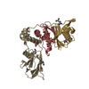
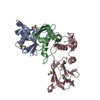
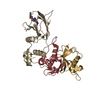
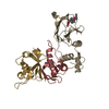
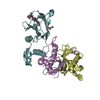
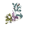
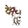
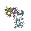
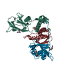
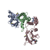
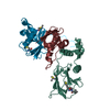
| Title | THE VHL-ELONGINC-ELONGINB STRUCTURE | ||||||
 Components Components |
| ||||||
 Keywords Keywords | TRANSCRIPTION / TUMOR SUPPRESSOR / CANCER / UBIQUITIN / BETA SANDWICH / TRANSCRIPTIONAL ELONGATION | ||||||
| Function / homology |  Function and homology information Function and homology informationregulation of cellular response to hypoxia / negative regulation of receptor signaling pathway via JAK-STAT / RHOBTB3 ATPase cycle / target-directed miRNA degradation / elongin complex / Replication of the SARS-CoV-1 genome / transcription elongation factor activity / VCB complex / Cul5-RING ubiquitin ligase complex / ubiquitin-dependent protein catabolic process via the C-end degron rule pathway ...regulation of cellular response to hypoxia / negative regulation of receptor signaling pathway via JAK-STAT / RHOBTB3 ATPase cycle / target-directed miRNA degradation / elongin complex / Replication of the SARS-CoV-1 genome / transcription elongation factor activity / VCB complex / Cul5-RING ubiquitin ligase complex / ubiquitin-dependent protein catabolic process via the C-end degron rule pathway / Cul2-RING ubiquitin ligase complex / SUMOylation of ubiquitinylation proteins / negative regulation of transcription elongation by RNA polymerase II / Pausing and recovery of Tat-mediated HIV elongation / Tat-mediated HIV elongation arrest and recovery / HIV elongation arrest and recovery / Pausing and recovery of HIV elongation / negative regulation of signal transduction / Tat-mediated elongation of the HIV-1 transcript / Formation of HIV-1 elongation complex containing HIV-1 Tat / Formation of HIV elongation complex in the absence of HIV Tat / ubiquitin-like ligase-substrate adaptor activity / RNA Polymerase II Transcription Elongation / Formation of RNA Pol II elongation complex / negative regulation of TORC1 signaling / RNA Polymerase II Pre-transcription Events / ciliary tip / transcription corepressor binding / negative regulation of autophagy / protein serine/threonine kinase binding / TP53 Regulates Transcription of DNA Repair Genes / positive regulation of cell differentiation / transcription initiation at RNA polymerase II promoter / transcription elongation by RNA polymerase II / Inactivation of CSF3 (G-CSF) signaling / Vif-mediated degradation of APOBEC3G / Evasion by RSV of host interferon responses / Oxygen-dependent proline hydroxylation of Hypoxia-inducible Factor Alpha / cell morphogenesis / Regulation of expression of SLITs and ROBOs / ubiquitin-protein transferase activity / transcription corepressor activity / positive regulation of proteasomal ubiquitin-dependent protein catabolic process / Antigen processing: Ubiquitination & Proteasome degradation / microtubule cytoskeleton / Neddylation / regulation of gene expression / protein-containing complex assembly / Replication of the SARS-CoV-2 genome / cellular response to hypoxia / DNA-binding transcription factor binding / amyloid fibril formation / molecular adaptor activity / ubiquitin-dependent protein catabolic process / proteasome-mediated ubiquitin-dependent protein catabolic process / protein-macromolecule adaptor activity / protein stabilization / cilium / protein ubiquitination / negative regulation of cell population proliferation / negative regulation of gene expression / ubiquitin protein ligase binding / regulation of transcription by RNA polymerase II / regulation of DNA-templated transcription / negative regulation of apoptotic process / positive regulation of DNA-templated transcription / enzyme binding / negative regulation of transcription by RNA polymerase II / endoplasmic reticulum / mitochondrion / proteolysis / nucleoplasm / nucleus / plasma membrane / cytosol Similarity search - Function | ||||||
| Biological species |  Homo sapiens (human) Homo sapiens (human) | ||||||
| Method |  X-RAY DIFFRACTION / SYNCHROTRON / OTHER / Resolution: 2.7 Å X-RAY DIFFRACTION / SYNCHROTRON / OTHER / Resolution: 2.7 Å | ||||||
 Authors Authors | Stebbins, C.E. / Kaelin, W.G. / Pavletich, N.P. | ||||||
 Citation Citation | Journal: Science / Year: 1999 Title: Structure of the VHL-ElonginC-ElonginB complex: implications for VHL tumor suppressor function. Authors: Stebbins, C.E. / Kaelin Jr., W.G. / Pavletich, N.P. | ||||||
| History |
|
- Structure visualization
| Structure viewer | Molecule: MolmilJmol/JSmol |
|---|
- Downloads & links
-Download
| PDBx/mmCIF format | 1vcb.cif.gz | 276 KB | Display | PDBx/mmCIF format |
|---|---|---|---|---|
| PDB format | pdb1vcb.ent.gz | 224.2 KB | Display | PDB format |
| PDBx/mmJSON format | 1vcb.json.gz | Tree view | PDBx/mmJSON format | |
| Others |  Other downloads Other downloads |
-Validation report
| Arichive directory | https://data.pdbj.org/pub/pdb/validation_reports/vc/1vcbftp://data.pdbj.org/pub/pdb/validation_reports/vc/1vcb | HTTPS FTP |
|---|
-Related structure data
| Similar structure data |
|---|


-Links
 PDBj
PDBj


- Assembly
| Deposited unit | 
| ||||||||||||||||||||||||||||||||||||||||||||||||||||
|---|---|---|---|---|---|---|---|---|---|---|---|---|---|---|---|---|---|---|---|---|---|---|---|---|---|---|---|---|---|---|---|---|---|---|---|---|---|---|---|---|---|---|---|---|---|---|---|---|---|---|---|---|---|
| 1 | 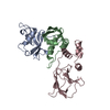
| ||||||||||||||||||||||||||||||||||||||||||||||||||||
| 2 | 
| ||||||||||||||||||||||||||||||||||||||||||||||||||||
| 3 | 
| ||||||||||||||||||||||||||||||||||||||||||||||||||||
| 4 | 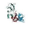
| ||||||||||||||||||||||||||||||||||||||||||||||||||||
| Unit cell |
| ||||||||||||||||||||||||||||||||||||||||||||||||||||
| Noncrystallographic symmetry (NCS) | NCS oper:
|
-Components
| #1: Protein | Mass: 13147.781 Da / Num. of mol.: 4 / Fragment: RESIDUES 1-120 Source method: isolated from a genetically manipulated source Details: DISORDERED RESIDUES: 99-120 / Source: (gene. exp.) Homo sapiens (human) / Plasmid: PGEX-4T3 / Species (production host): Escherichia coli / Gene (production host): VHL / Production host:  #2: Protein | Mass: 12485.135 Da / Num. of mol.: 4 / Fragment: RESIDUES 17-112 Source method: isolated from a genetically manipulated source Details: DISORDERED RESIDUES: 50-57 / Source: (gene. exp.) Homo sapiens (human) / Plasmid: PBB75 / Species (production host): Escherichia coli / Gene (production host): VHL / Production host: #3: Protein | Mass: 18558.162 Da / Num. of mol.: 4 / Fragment: RESIDUES 54-213 Source method: isolated from a genetically manipulated source Details: DISORDERED RESIDUES: 54-62, 205-213 / Source: (gene. exp.) Homo sapiens (human) / Description: VHL(54-213) ALTERNATIVE ENDOGENOUS POLYPEPTIDE / Plasmid: PGEX-4T3 / Species (production host): Escherichia coli / Gene (production host): VHL / Production host: #4: Water | ChemComp-HOH / |  Mass: 18.015 Da / Num. of mol.: 454 / Source method: isolated from a natural source / Formula: H2O Mass: 18.015 Da / Num. of mol.: 454 / Source method: isolated from a natural source / Formula: H2O |
|---|
-Experimental details
-Experiment
| Experiment | Method: X-RAY DIFFRACTION / Number of used crystals: 1 |
|---|
- Sample preparation
| Crystal | Density Matthews: 2.24 Å3/Da / Density % sol: 43 % | ||||||||||||||||||||||||||||||
|---|---|---|---|---|---|---|---|---|---|---|---|---|---|---|---|---|---|---|---|---|---|---|---|---|---|---|---|---|---|---|---|
| Crystal grow | pH: 5.7 Details: 10-15% PEG 2000, 200MM MAGNESIUM ACETATE, 100MM SODIUM CACODYLATE PH 5.7 | ||||||||||||||||||||||||||||||
| Crystal | *PLUS | ||||||||||||||||||||||||||||||
| Crystal grow | *PLUS Temperature: 4 ℃ / Method: vapor diffusion, hanging drop | ||||||||||||||||||||||||||||||
| Components of the solutions | *PLUS
|
-Data collection
| Diffraction | Mean temperature: 113 K |
|---|---|
| Diffraction source | Source: SYNCHROTRON / Site: CHESS  / Beamline: F1 / Wavelength: 0.918 / Beamline: F1 / Wavelength: 0.918 |
| Detector | Type: ADSC QUANTUM 4 / Detector: CCD / Date: Sep 20, 1998 |
| Radiation | Protocol: SINGLE WAVELENGTH / Monochromatic (M) / Laue (L): M / Scattering type: x-ray |
| Radiation wavelength | Wavelength: 0.918 Å / Relative weight: 1 |
| Reflection | Resolution: 2.7→20 Å / Num. obs: 41219 / % possible obs: 90.9 % / Observed criterion σ(I): 2 / Redundancy: 3 % / Rmerge(I) obs: 0.07 / Rsym value: 7 |
| Reflection | *PLUS Num. measured all: 123622 / Rmerge(I) obs: 0.07 |
- Processing
| Software |
| ||||||||||||||||||||||||||||||||||||||||||||||||||||||||||||
|---|---|---|---|---|---|---|---|---|---|---|---|---|---|---|---|---|---|---|---|---|---|---|---|---|---|---|---|---|---|---|---|---|---|---|---|---|---|---|---|---|---|---|---|---|---|---|---|---|---|---|---|---|---|---|---|---|---|---|---|---|---|
| Refinement | Method to determine structure: OTHER / Resolution: 2.7→20 Å / Cross valid method: THROUGHOUT / σ(F): 2
| ||||||||||||||||||||||||||||||||||||||||||||||||||||||||||||
| Solvent computation | Solvent model: FLAT MODEL | ||||||||||||||||||||||||||||||||||||||||||||||||||||||||||||
| Displacement parameters | Biso mean: 54.1 Å2 | ||||||||||||||||||||||||||||||||||||||||||||||||||||||||||||
| Refinement step | Cycle: LAST / Resolution: 2.7→20 Å
| ||||||||||||||||||||||||||||||||||||||||||||||||||||||||||||
| Refine LS restraints |
| ||||||||||||||||||||||||||||||||||||||||||||||||||||||||||||
| Refine LS restraints NCS | NCS model details: RESTRAINTS (500KCAL MOL^-1 ANGSTROM^-2) | ||||||||||||||||||||||||||||||||||||||||||||||||||||||||||||
| Software | *PLUS Name: CNS / Version: 0.3 / Classification: refinement | ||||||||||||||||||||||||||||||||||||||||||||||||||||||||||||
| Refinement | *PLUS Highest resolution: 2.7 Å / Lowest resolution: 20 Å / σ(F): 2 / % reflection Rfree: 5 % / Rfactor obs: 0.239 | ||||||||||||||||||||||||||||||||||||||||||||||||||||||||||||
| Solvent computation | *PLUS | ||||||||||||||||||||||||||||||||||||||||||||||||||||||||||||
| Displacement parameters | *PLUS Biso mean: 54.1 Å2 | ||||||||||||||||||||||||||||||||||||||||||||||||||||||||||||
| Refine LS restraints | *PLUS
|
