 Movie
Movie Controller


+ Open data

- Basic information
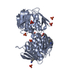
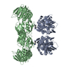
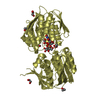
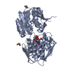
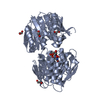
| Entry | Database: PDB / ID: 1ejc | ||||||
|---|---|---|---|---|---|---|---|
| Title | Crystal structure of unliganded mura (type2) | ||||||
 Components Components | UDP-N-ACETYLGLUCOSAMINE ENOLPYRUVYLTRANSFERASE | ||||||
 Keywords Keywords | TRANSFERASE / inside-out alpha/beta barrel | ||||||
| Function / homology |  Function and homology information Function and homology informationUDP-N-acetylglucosamine 1-carboxyvinyltransferase activity / UDP-N-acetylgalactosamine biosynthetic process / UDP-N-acetylglucosamine 1-carboxyvinyltransferase / peptidoglycan biosynthetic process / cell wall organization / regulation of cell shape / cell division / cytoplasm Similarity search - Function | ||||||
| Biological species |  Enterobacter cloacae (bacteria) Enterobacter cloacae (bacteria) | ||||||
| Method |  X-RAY DIFFRACTION / Resolution: 1.8 Å X-RAY DIFFRACTION / Resolution: 1.8 Å | ||||||
 Authors Authors | Eschenburg, S. / Schonbrunn, E. | ||||||
 Citation Citation | Journal: Proteins / Year: 2000 Title: Comparative X-ray analysis of the un-liganded fosfomycin-target murA. Authors: Eschenburg, S. / Schonbrunn, E. #1: Journal: Biochemistry / Year: 2000Title: Role of the Loop Containing Residue 115 in the Induced-fit Mechanism of the Bacterial Cell Wall Biosynthetic Enzyme MurA Authors: Schonbrunn, E. / Eschenburg, S. / Krekel, F. / Luger, K. / Amrhein, N. #2: Journal: Structure / Year: 1996Title: Crystal Structure of UDP-N-acetylglucosamine Enolpyruvyltransferase, the Target of the Antibiotic Fosfomycin Authors: Schonbrunn, E. / Sack, S. / Eschenburg, S. / Perrakis, A. / Krekel, F. / Amrhein, N. / Mandelkow, E. | ||||||
| History |
|
- Structure visualization
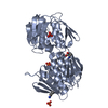
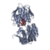
| Structure viewer | Molecule: MolmilJmol/JSmol |
|---|
- Downloads & links
-Download
| PDBx/mmCIF format | 1ejc.cif.gz | 107.2 KB | Display | PDBx/mmCIF format |
|---|---|---|---|---|
| PDB format | pdb1ejc.ent.gz | 80.4 KB | Display | PDB format |
| PDBx/mmJSON format | 1ejc.json.gz | Tree view | PDBx/mmJSON format | |
| Others |  Other downloads Other downloads |
-Validation report
| Arichive directory | https://data.pdbj.org/pub/pdb/validation_reports/ej/1ejcftp://data.pdbj.org/pub/pdb/validation_reports/ej/1ejc | HTTPS FTP |
|---|
-Related structure data


-Links
 PDBj
PDBj
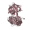
- Assembly
| Deposited unit | 
| ||||||||
|---|---|---|---|---|---|---|---|---|---|
| 1 |
| ||||||||
| Unit cell |
|
-Components
| #1: Protein | Mass: 44829.406 Da / Num. of mol.: 1 Source method: isolated from a genetically manipulated source Source: (gene. exp.) Enterobacter cloacae (bacteria) / Production host: References: UniProt: P33038, UDP-N-acetylglucosamine 1-carboxyvinyltransferase | ||||
|---|---|---|---|---|---|
| #2: Chemical | ChemComp-PO4 /   Mass: 94.971 Da / Num. of mol.: 1 / Source method: obtained synthetically / Formula: PO4 Mass: 94.971 Da / Num. of mol.: 1 / Source method: obtained synthetically / Formula: PO4 | ||||
| #3: Chemical | ChemComp-GOL /   Mass: 92.094 Da / Num. of mol.: 4 / Source method: obtained synthetically / Formula: C3H8O3 Mass: 92.094 Da / Num. of mol.: 4 / Source method: obtained synthetically / Formula: C3H8O3#4: Water | ChemComp-HOH / |  Mass: 18.015 Da / Num. of mol.: 534 / Source method: isolated from a natural source / Formula: H2O Mass: 18.015 Da / Num. of mol.: 534 / Source method: isolated from a natural source / Formula: H2OHas protein modification | Y | |
-Experimental details
-Experiment
| Experiment | Method: X-RAY DIFFRACTION / Number of used crystals: 1 |
|---|
- Sample preparation
| Crystal | Density Matthews: 2.15 Å3/Da / Density % sol: 42.89 % | |||||||||||||||||||||||||
|---|---|---|---|---|---|---|---|---|---|---|---|---|---|---|---|---|---|---|---|---|---|---|---|---|---|---|
| Crystal grow | Temperature: 292 K / Method: vapor diffusion, hanging drop / pH: 6.5 Details: 10 % PEG 20000, 0.1 M MES, pH 6.5, VAPOR DIFFUSION, HANGING DROP, temperature 292K | |||||||||||||||||||||||||
| Crystal grow | *PLUS Temperature: 19 ℃ | |||||||||||||||||||||||||
| Components of the solutions | *PLUS
|
-Data collection
| Diffraction | Mean temperature: 120 K |
|---|---|
| Diffraction source | Source: ROTATING ANODE / Type: RIGAKU RU300 / Wavelength: 1.5418 |
| Detector | Type: RIGAKU RAXIS IV / Detector: IMAGE PLATE / Date: May 5, 1999 |
| Radiation | Protocol: SINGLE WAVELENGTH / Monochromatic (M) / Laue (L): M / Scattering type: x-ray |
| Radiation wavelength | Wavelength: 1.5418 Å / Relative weight: 1 |
| Reflection | Resolution: 1.8→20 Å / Num. all: 36451 / Num. obs: 36451 / % possible obs: 99.6 % / Observed criterion σ(F): 0 / Observed criterion σ(I): 0 / Redundancy: 6.8 % / Biso Wilson estimate: 17.3 Å2 / Rmerge(I) obs: 0.046 / Net I/σ(I): 28.4 |
| Reflection shell | Resolution: 1.8→1.84 Å / Rmerge(I) obs: 0.225 / % possible all: 99.2 |
| Reflection | *PLUS Num. measured all: 251049 |
| Reflection shell | *PLUS % possible obs: 99.2 % / Mean I/σ(I) obs: 5.8 |
- Processing
| Software |
| |||||||||||||||||||||||||
|---|---|---|---|---|---|---|---|---|---|---|---|---|---|---|---|---|---|---|---|---|---|---|---|---|---|---|
| Refinement | Resolution: 1.8→20 Å / Rfactor Rfree error: 0.006 / Isotropic thermal model: restrained / Cross valid method: THROUGHOUT / σ(F): 0 / σ(I): 0 / Stereochemistry target values: Engh & Huber Details: HOH 1 occupies an alternate position of alternate conformation A NH1 ARG 295. HOH 1 has alternate position A.
| |||||||||||||||||||||||||
| Displacement parameters | Biso mean: 19 Å2
| |||||||||||||||||||||||||
| Refine analyze |
| |||||||||||||||||||||||||
| Refinement step | Cycle: LAST / Resolution: 1.8→20 Å
| |||||||||||||||||||||||||
| Refine LS restraints |
| |||||||||||||||||||||||||
| Refine LS restraints NCS | NCS model details: NONE | |||||||||||||||||||||||||
| LS refinement shell | Resolution: 1.8→1.91 Å / Rfactor Rfree error: 0.022 / Total num. of bins used: 6
| |||||||||||||||||||||||||
| Software | *PLUS Name: CNS / Classification: refinement | |||||||||||||||||||||||||
| Refine LS restraints | *PLUS
|
